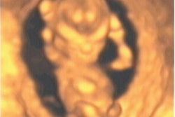
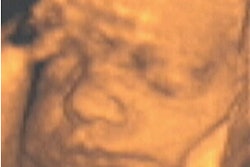
Combining transperineal grayscale and color Doppler ultrasound yields increased detection and comprehensive characterization of perianal abscesses and fistulas, according to research published in the January issue of the Journal of Ultrasound in Medicine.
"Combining color Doppler and grayscale sonography significantly augmented the operator’s diagnostic confidence," wrote researchers from the departments of radiology and surgery at Innsbruck University Hospital in Austria. "This could have a considerable clinical effect because transperineal sonography may spare patients painful clinical examinations and may support noninvasive treatment planning for patients with perianal inflammatory disease."
One of two senior radiologists on the Innsbruck University study team performed both types of ultrasound studies on 87 patients with suspected perianal inflammatory disorders between January 2000 and December 2002 (JUM, January 2004, Vol. 23:1, pp. 19-27).
Using a linear 4-to-7 MHz transducer on an HDI 5000 system (Philips Medical Systems, Andover, MA), the entire perianal region was scanned for the detection of suspected inflammatory disorders; each detected inflammatory disorder was evaluated to determine its morphologic characteristics and extent.
Color Doppler sonography was applied to assess the presence of increased vascularity in the perianal region. The two radiologists arrived at their diagnoses by consensus.
To compare results with the surgical findings, the researchers then performed a receiver operating characteristics (ROC) analysis for lesion detection and the Spearman test for lesion characterization. They also performed logistic regression analysis to assess whether increased perineal vascularity was a predictive factor of perianal inflammatory disease.
Of the 87 patients, 62 had 77 confirmed perianal inflammatory disorder cases. Grayscale sonography turned in a "significantly good performance" in the detection (area under the ROC curve = 0.86) and characterization (r = 0.65) of perianal inflammatory disease. Sensitivity for detection of perianal fistulas and abscesses was 100% and 94%, respectively.
Adding color Doppler ultrasound increased diagnostic confidence slightly (area under the curve = 0.89), but significantly, according to the researchers. From logistic regression analysis, the study team concluded that hypervascularity at the periphery of a perianal lesion is a significant independent predictor of an inflammatory disease.
The researchers noted that transperineal sonography is a fast, noninvasive, and painless technique for evaluating the structures of the pelvic floor, the diseases of which, particularly perianal abscesses and fistulas, are often accompanied by severe pain. As digital rectal examination, rectoscopy, and transrectal sonography may provoke pain and might not be tolerated by patients, patients frequently are subjected to examinations under general anesthesia and may undergo surgical exploration for future therapeutic planning, the authors said.
"In this study, we found that transperineal sonography can provide valuable information about the perineum and perianal structures in a relatively short time and with less discomfort to the patient," the authors wrote.
The study team also noted that the location, extension, and topographic relationship between a perianal abscess and the rectum, the prostate in men, and the vagina in women could be determined precisely. Also, perianal fistulas -- even bifurcated, complex, and blind-ended fistulas -- could be mapped from the external to the internal openings, including their entire extension.
Because all segments of an abscess or fistula are preoperatively recognized by the surgeon, transperineal sonography facilitates acute surgical intervention, leading to resection of the entire inflammatory lesion and thus reducing the risk of disease recurrence, according to the authors. The ultrasound technique also offers the ability to visualize and locate the internal opening of a fistula, which aids surgeons in finding the ostium during surgery.
In comparison with endoanal sonography, transperineal sonography adds information about the entire pelvic floor, particularly when panoramic imaging is applied, according to the researchers. This added capability provides a better idea of the presence or absence of other painful lesions in the pelvic floor, which can help in deciding for or against a surgical intervention.
"Surgical exploration could be avoided in patients with perineal fistulas that are not accompanied by abscesses, and initial conservative therapy with anti-inflammatory agents, antibiotics, and enemas could be warranted," the authors wrote. "According to the results of our study, the high specificity and negative predictive value of transperineal sonography in the detection of perianal abscesses indicate its accuracy in excluding abscesses."
MRI is considered the examination of choice for evaluating anorectal disease, owing to its comprehensive perceptibility of the perianal region and high accuracy in detection of perianal fistulas. However, MRI is expensive and may not always be as readily available as sonography, the authors said.
In addition, endorectal MRI is affected by its limited field of view and patient discomfort; it also requires contrast injection, which is not needed for transperineal sonography, the researchers said. Color Doppler sonography tended to show the pathologic vasculature of perianal abscesses and fistulas, they noted.
"In this study, color Doppler sonography augmented the recognition of acute perianal inflammatory conditions in three patients with acute fissures despite missing the fissures themselves," the authors wrote. "Although color Doppler sonography did not improve the sensitivity and specificity for detecting perianal abscesses and fistulas, the presence of pathologic vascular structures in the peripheries of the abscesses and fistulas supported the findings shown on grayscale sonography, and thus improved the operator’s diagnostic confidence."
The typical peripheral hypervascularization of abscesses also may allow differentiation between an abscess and a solid perianal tumor, which tends to show central hypervascularization, they noted.
By Erik L. RidleyAuntMinnie.com staff writer
January 15, 2004
Related Reading
Focused ultrasound shows promise as treatment for advanced renal cancer, December 16, 2003
3-D ultrasound shows promise in neonatal, pediatric neurosonography, November 4, 2003
No additional info offered by 3-D sonography of adnexal masses, June 19, 2003
3-D ultrasound offers reliable assessment of gallbladder, bile ducts, May 5, 2003
Power Doppler US spots tumor vascularity for prostate cancer staging, April 30, 2003
Copyright © 2004 AuntMinnie.com