For decades, imaging specialists have sought a solution to a very common diagnostic dilemma: When a patient presents with severe abdominal pain, how can one tell if it’s acute appendicitis?
This dilemma occurs not only frequently, but often urgently. And the wrong answer can result in a ruptured appendix or an unnecessary surgery. Hence, a number of researchers have explored the diagnostic potential of both CT and ultrasound, with both modalities achieving impressive accuracy in certain studies.
But the most impressive results may be unattainable for most medical facilities, according to a new study out of St. Elizabeth Hospital in Tilburg, the Netherlands. (American Journal of Roentgenology, November 2003, Vol. 181:5, pp.1355-1359).
Members of the hospital’s surgery, radiology, and pathology staff worked together with statisticians in Amsterdam to conduct a particularly rigorous comparison of CT versus ultrasound in a "realistic clinical setting" -- i.e., where both body imaging specialists and general radiology staff members were reading the potential appendicitis exams.
"As far as we know, this study is the first blinded prospective study comparing the reliability of CT without administration of any form of enteric contrast material (unenhanced CT) and sonography for diagnosing acute appendicitis in daily practice in an unselected population of patients admitted to a general community teaching hospital with suspected acute appendicitis," the authors wrote.
The only comparable study cited by the researchers was one that compared sonography and contrast-enhanced CT in unselected patients at a university hospital with radiologists of widely varying experience The researchers found that CT performed significantly better than sonography, but also found interobserver variability in CT evaluation (AJR, April 2001, Vol. 176:4, pp. 933-941).
The Tilburg researchers opted to do CT without contrast, based on several studies that found unenhanced scans to be just as accurate for the detection of acute appendicitis.
For nearly two years, from August 1998 to June 2000, all patients who showed up at St. Elizabeth’s with suspected appendicitis were evaluated by a senior surgical resident or staff surgeon. Sixty patients were so ill that they underwent immediate surgery and were excluded from the imaging study.
Twelve patients declined to participate in the study, and another 10 were dropped due to "logistic problems" in radiology. Surgical staff either forgot or refused to include another 14 patients in the study, and nine were excluded because the surgeon disagreed with the general practitioner’s diagnosis of possible appendicitis. The remaining 234 patients underwent both imaging exams.
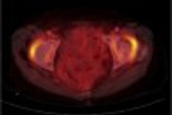
Single-breathhold CT images from the L2 vertebra to the pubic symphysis were taken with a single-detector helical scanner (Tomoscan AV, Philips Medical Systems, Andover, MA), using 5-mm beam collimation and 5-mm/sec table speed (pitch of 1, 120 kVp, 100-250 mAs). An enlarged appendix of 6 mm in diameter or greater was deemed positive for acute appendicitis.
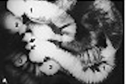
Ultrasound exams were performed with 5-12 MHz linear array, 5-2 MHz curved array, and 8-5 MHz curved array transducers (HDI 3000, ATL-Philips Medical Systems), using the graded compression technique (Radiology,1986, Vol. 158, pp. 255-360).
"On sonography, the primary criterion to establish the diagnosis of acute appendicitis was direct visualization of the inflamed appendix: a concentrically layered, small, sausage-like structure found at the point of tenderness," the Tilburg researchers wrote.
The imaging exams were performed within one hour by two radiologists blinded to the other’s findings. Just two of the 12 participating radiologists were body imaging specialists. Surgeons were not informed of the radiologic diagnoses prior to operating, although in eight cases the radiologists informed an independent surgeon about findings such as diverticulitis that would significantly affect patient management.
Of the 226 patients completing the study protocol, 199 underwent surgery and 66% of those were found to have appendicitis. In 41 of the remaining 67 patients, another diagnosis was made, including cholecystitis, cecal tumors, and ovarian cysts. Most of these non-appendicitis findings were seen on both CT and US.
Yet obviously, neither CT nor US performed impeccably in definitively diagnosing appendicitis, and, in fact, performed worse than in many other published studies. Early research had shown sensitivity of 70%-100% for helical CT and 75%-90% for sonography, and specificity of 91%-99% and 86%-100% respectively.
At St. Elizabeth’s, CT demonstrated 76% sensitivity, 83% specificity, 90% positive predictive value, and 64% negative predictive value. Ultrasound achieved very similar results on the same analyses, with 79%, 78%, 87%, and 65% respectively.
Both modalities achieved an overall accuracy of 78% -- a particularly damning result for CT, given that it takes more effort, time, and money to perform than ultrasound. But the Tilburg group was unapologetic, suggesting their results were more realistic than the earlier studies by research-oriented academic centers.
"The disappointing CT results in this study are more likely to reflect the CT performance in an average hospital," they wrote. Moreover, the study design also required their radiologists to address one of the ongoing customer complaints about diagnostic imaging.
"Another reason for the lower accuracy of CT in our study may be the exclusion of the possibility for equivocal test results by forcing the radiologist to make a decision whether or not acute appendicitis was present," the authors wrote. "In daily practice our surgical staff demands a clear statement by the radiologists."
"In our opinion, the introduction of CT and sonography as a standard procedure in the workup of acute appendicitis can be worthwhile only if the surgeon can rely fully on CT and sonography performed in the hospital," the researchers concluded. "If a small risk of a perforated acute appendicitis is still present even when both CT and sonography show a normal appendix, most surgeons will neglect the benefits of these additional radiologic tools."
By Tracie L. ThompsonAuntMinnie.com staff writer
December 10, 2003
Related Reading
Pre-operative CT best suited for atypical cases of kiddie appendicitis, August 1, 2003
CT overtakes ultrasound for pediatric acute appendicitis, May 9, 2003
US plus CT improves pediatric appendicitis management, March 25, 2003
Ultrasound differentiates causes of abdominal pain in kids, October 4, 2002
Copyright © 2003 AuntMinnie.com