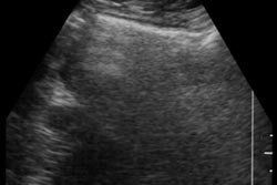
(Ultrasound Review) Often tubal pathology is misdiagnosed as ovarian in origin. The two most reliable signs for identifying tubal pathology are the elongated shape and the presence of septa of the tubal wall.
In this study of 25 women with fallopian tube masses, grayscale and color Doppler were used to characterize lesions and possibly differentiate between acute and chronic tubal disease. The study was published in the Journal of Ultrasound in Medicine.
There were 18 women with hydrosalpinx, one with a tubo-ovarian abscess, and one with a tubal torsion. Findings were confirmed at laparoscopy, laparotomy, hysterosalpingography, or abscess drainage.
Transvaginal ultrasound imaging was performed using a 4-7-MHz or 6.5-MHz transducer. Color Doppler parameters were adjusted for maximum sensitivity to low flow. Pulsed-wave Doppler was used to obtain velocity waveforms from the mass, and the resistive index (RI) was calculated.
Ultrasound characteristics of hydrosalpinx included elongated cystic lesion in the adnexa pouch of Douglas with thin borders, clear fluid, and an RI of 0.75. In contrast, a tubo-ovarian abscess showed thick borders with turbid fluid and a mean RI of 0.45.
For abscesses, the abundant vessels had high flow, with a lower vascular resistance. This is typically found with inflammatory conditions.
Color Doppler representing wall vasculature was demonstrated in all cases, with the exception of the tubal torsion, and there was a significant difference in RI comparing acute and chronic tubal conditions.
"The addition of color Doppler flow sonography to the investigation of chronic pelvic pain can distinguish between hydrosalpinx and dilated veins, as is the case with pelvic congestion syndrome," the authors said. "The addition of color Doppler flow sonography in investigating the suspected tubal lesions may reduce the false-positive cases."
The authors conclude that color and pulsed Doppler should be used whenever a tubal abnormality is demonstrated to further characterize the type of lesion.
J Ultrasound Med 2000; 19:645–649
Yaron Zalel et al
Ultrasound Unit, Obstetrics and Gynecology Dept, Sheba Medical Center, Tel Hashomer 52621, Israel
By Ultrasound Review
December 29, 2000
Copyright © 2000 AuntMinnie.com