Brain-volume changes on MRI can be a reliable biomarker for the neuropathology of Alzheimer’s disease, according to a study out of the Oregon Health and Sciences University in Portland. Specifically, the researchers looked at whether the accumulation of senile plaques (SP) and neurofibrillary tangles (NFT) can be used to monitor disease progression in clinical trials.
"The first objective of this study was to confirm that the degree of brain-volume atrophy observed prior to death by MRI volumetrics is predictive of subsequent (Alzheimer’s disease) neuropathology ... second, we sought to determine which specific brain regions were best able to predict pathology on the basis of volume measurement," wrote lead author Dr. Lisa Silbert and colleagues, some of whom are also associated with the Portland Veterans Affairs Medical Center (Neurology, August 26, 2003, Vol. 61:4, pp. 487-492).
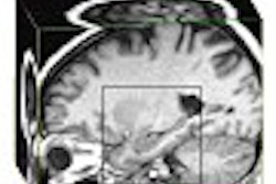
For this study, 39 elderly subjects (15 healthy controls, 24 with cognitive impairment) were followed for a mean of 5.8 years. They’d been selected on the basis of having had at least one MR scan prior to autopsy and, for those with dementia, a diagnosis of probable Alzheimer’s disease. Images were acquired on a 1.5-tesla MR system. The protocol included continuous-slice, multiecho, multiplanar image acquisition with 4-mm thick coronal slices. T1-weighted midsagittal images also were acquired.
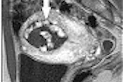
Four areas comprised the region of interest (ROI): total supratentorial brain volume, total ventricular volume, temporal horn volume, and hippocampal body volume. The median postmortem interval was 12.3 hours, with the preserved brain examined by a neuropathologist who had no knowledge of the MR data.
According to the results, in cognitively impaired subjects, the rate of ventricular volume increase over time was 5.5 cc/year, while the rate of central nervous system (CNS) volume decrease was -17.9 cc/year. In comparison, non-demented individuals showed a 3.3 cc/year increase in ventricular volume size and a -1.8 cc/year decrease in CNS volume.
In an evaluation of the relationship of the last MRI volumes to cortical neuropathology, the group found that a connection between the amount of cortical NFT burden and the total ventricular cerebrospinal fluid (CSF) volume, as well as the last total brain volume. They also found a relationship between plaque burden and last total ventricular volume prior to death in the cortical SP. Finally, they found a relationship between the degree of hippocampal NFT burden and total hippocampal volume prior to death in the subjects with cognitive impairment.
In summary, they determined that cortical NFT was the key to judging Alzheimer’s disease pathology: total brain volume change over time was related to the accumulation of cortical NFT; the rate of ventricular CSF volume increase was related to cortical NFT and SP; and the last hippocampal volume prior to death was related to hippocampal NFT burden. In addition, the rate of total ventricular CSF volume increase was the best predictor of cortical NFT and SP.
"The rate of ventricular volume enlargement can be used to monitor disease progression or response to treatment in future clinical trials that are targeted at NFT and SP pathology," the authors wrote. "As MRI hippocampal volumes taken at the time of death represent a point of maximal atrophy, they are likely to be more sensitive to reflecting underlying (Alzheimer’s disease) pathology."
By Shalmali PalAuntMinnie.com staff writer
September 5, 2003
Related Reading
Scans suggest why education prevents Alzheimer's, August 8, 2003
MR study shows antidepressants shield hippocampus from deterioration, August 5, 2003
Alzheimer's brains show features of immaturity, May 8, 2003
Imaging's role still evolving in dementia diagnosis, April 29, 2003
Copyright © 2003 AuntMinnie.com