SAN DIEGO - Patient preparation and image acquisition techniques can maximize the benefit of using FDG-PET for tumor diagnosis and staging. At the University of Texas, M.D. Anderson Cancer Center in Houston, the modality has been used to image patients with non-small cell lung cancer, lymphoma, melanoma, and colorectal, esophageal, breast, and head and neck cancer. Physicians at the center use FDG-PET to stage or identify tumors as well as to distinguish recurrence from post-treatment changes and to monitor response to therapy.
"One of the most important things you can do for quality PET scans is to have consistency of technique in acquisition and patient preparation," said Dr. Homer Macapinlac of the division of diagnostic imaging, department of nuclear medicine PET section at M.D. Anderson Cancer Center. Macapinlac spoke at the American Roentgen Ray Society meeting on Sunday.
Preparation
Patients scheduled for PET imaging are asked to fast for a minimum of six hours (the preferred length of time is overnight) before their scan. At the meal before their fast, patients are requested to eat foods high in protein and low in carbohydrates to minimize myocardial activity, Macapinlac said.
Diabetic patients are asked to take their medication and eat a normal breakfast at an early hour. The center schedules its diabetic patients for imaging at midday. "We’ve got solid protocols in place for FDG-PET scans of diabetics," Macapinlac noted.
Patients who are nervous or are known to have post-therapeutic neuromuscular deficiencies or muscle spasms are given alprazolam (Xanax, Pharmacia and Upjohn) approximately 15-30 minutes before the FDG injection. According to Macapinlac, this minimizes the uptake by normal skeletal muscle, which would make neck and supraclavicular node evaluation difficult.
"Our protocol for FDG-PET scans for staging patients with head, neck, or breast cancer is to give them Xanax orally before FDG injection, and ask them to come to the imaging session with someone who can drive them home after we’re done," Macapinlac said.
All patients are encouraged to be well hydrated by drinking lots of water before the FDG injection. This enhances the flushing of the radiopharmaceutical from the urinary collecting system, which is the main route of excretion. Patients are injected with 10-20 mCi of FDG, which is then followed with a 10-mL saline flush.
After the FDG is administered, the center asks its patients to stay quiet and remain in a sitting or recumbent position for at least one hour. Those patients who are being imaged for cancers with a relatively low FDG uptake, such as breast, pancreas, or hepatocellular carcinoma, are asked to remain at rest for at least 90 minutes. Immediately before the scan, patients will be asked to empty their bladder and also drink water to remove any saliva retained in the mouth or esophagus, said Macapinlac.
Acquisition
The M.D. Anderson Cancer Center has two dedicated PET scanners, one manufactured by Siemens Medical Solutions (Malvern, PA), and the other by GE Medical Systems (Waukesha, WI). In addition, the department has also started acquiring images on a Discovery ST PET-CT unit (GE Medical Systems) that can perform eight-slice multidetector CT scans.
Images are acquired at emission in 2-D, and three-minute transmission scans are sequentially acquired for attenuation correction. When CT scans are acquired prior to PET scans, the center uses a protocol of 140 kVp and 140 mAs at a 15-mm rotation. Images are acquired at mid-expiration by the patient, and without IV or oral contrast material.
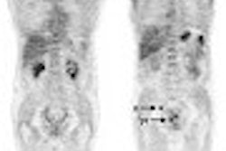
A PET scan at the center usually encompasses the skull base by starting at the top of the patient’s ears, and then covers the thorax and abdomen to the proximal femurs. For patients who are being staged or monitored for melanoma, the whole body is scanned.
"Patients are usually positioned on the gantry with their arms up for the PET examination, except those patients who are being scanned for melanoma, head and neck carcinoma, and patients with arm lesions, such as sarcomas," Macapinlac said.
Dedicated PET images are reconstructed by the center using vendor-supplied software that performs ordered subset expectation maximization, iterative reconstruction, and segmented attenuation correction, as well as standardized uptake value (SUV) formulas.
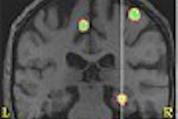
Brain imaging for tumors uses a slightly modified protocol of 5-10 mCi of FDG with an uptake phase of 30-45 minutes. The patients are imaged in a darkened room to minimize visual cortex activity and are kept in a quiet environment prior to scanning to minimize unilateral temporal lobe uptake of the radiotracer.
Coregistration scanning is usually performed on an MR scanner, rather than using CT imaging. The PET scans are performed in 3-D mode with calculated attenuation correction, Macapinlac said.
"Although PET is very useful, don’t forget that CT, MR, and x-ray are equally important in making an accurate diagnosis," he said. "Combined together, these modalities are incredibly powerful tools for diagnosing, staging, and monitoring tumors."
By Jonathan S. Batchelor
AuntMinnie.com staff writer
May 5, 2003
Related Reading
PET/CT growth sparks gains in U.S. PET market, April 15, 2003
German radiologists assess performance of PET-CT, April 10, 2003
PET/CT boosts confidence in rectal cancer staging, US yields high sensitivity, April 3, 2003
New protocol improves prognosis for advanced head/neck cancer, February 25, 2003
Studies illustrate benefits of FDG-PET, pre-planning in radiotherapy, January 27, 2003
Copyright © 2003 AuntMinnie.com