A novel hardware-coregistered PET-CT system improves the diagnosis of cancer significantly compared with PET alone, according to researchers at the University Hospital Zurich in Switzerland. The group's preliminary results also showed that adding PET improved diagnostic accuracy far more effectively than raising the CT dose did.
Over the past several years, a number of studies have shown that software-based coregistration of PET and CT images is more accurate than CT alone, especially for detecting non-small cell lung cancers and mediastinal lymph node metastases, the authors wrote in Radiology Online (September 19, 2002).
In 2000, a group from the University of Pittsburgh demonstrated the feasibility of a hardware-based coregistration system using a PET system combined with single-detector-row scanner (Clinical Positron Imaging, July 2000, Vol. 3:4, p. 161).
The current study, combining a PET with a multidetector-row CT scanner, was supported in part by the prototype system's manufacturer, GE Medical Systems of Waukesha, WI.
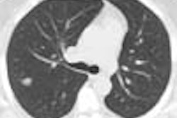
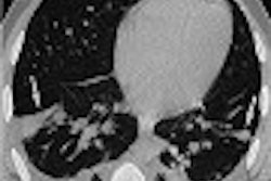
The group examined 53 consecutive patients, 25 men and 18 women aged 39-84 (mean age 61), all with diagnosed or suspected malignancies. Images were acquired using a prototype PET-CT inline system that combined GE's Discovery LS CT scanner and Advance NXi PET scanner. The axes of both systems were aligned to coincide mechanically, and the same table was used to acquire both kinds of images by advancing automatically from one modality to the other, the authors wrote.
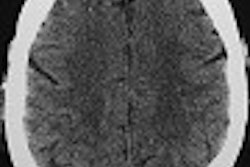
Following a four-hour fast, patients were injected with 370 MBq of FDG, with PET acquisition beginning 45 minutes later, after CT scanning. The group acquired 6 contiguous volumes of 14.6 cm each from the cerebellum to the pelvic floor.
Four CT scans were acquired of each patient using increasing mAs of 10, 40, 80 and 120, with other parameters remaining constant: 0.5 per second resolution, 140 kV, section pitch 6, 5 mm reconstructed section thickness. Shallow breathing was allowed during CT acquisition.
After CT scanning, the table advanced automatically into the PET gantry. The group acquired 35 two-dimensional non-attenuation-corrected scans in each of six incremental table positions covering 867 mm. Total PET acquisition time was 42 minutes.
PET images were first analyzed alone, then fused with CT images acquired at increasingly higher mA settings: 10, 40, 80 and 120 mA. Staging was also performed in an effort to assess the clinical effect of PET-CT examinations.
"Since lesion classification and location were taken to be relevant for correct diagnosis in malignant lesions, the clinical or pathological stage was used as the standard of reference," the authors wrote. "The clinical stage was determined on the basis of histologic proof of the primary lesion and of crucial secondary lesions. In cases of extended disease, no further workup was performed. The pathologic stage was determined on the basis of findings in macro- and micropathologic specimens obtained at surgery with curative intent."
Specificity and sensitivity of the technique were calculated based on lesion-by-lesion analysis in addition to the standard of reference, the authors wrote. Overall, 287 lesions were analyzed.
Using PET alone, 137 lesions were regarded as tumors, 40 as inflammatory changes, and 44 as other lesions. Compared with the reference standard, 123/137 were true positives (TP) six were false-positives (FP), and 8 false negatives (FN). Following exclusion of 61 undecided lesions, PET had a sensitivity of 90% and specificity of 93% compared with the reference standard.
PET combined with 10-mA CT defined 135 lesions as tumors, 50 as inflammation, and 54, other. Compared with the reference standard, 133 lesions were TP, 109 TN, and 5 FN, the authors wrote. Compared with the reference standard, low-dose combined technique yielded sensitivity of 96% and specificity of 99%.
"The location was changed for 12 of the 22 lesions, which became TP rather than TN," the authors wrote. "The undecided lesions were reduced (from 61) to 39."
PET plus 40-mA CT classified the same 135 lesions as tumors, while changing 3 of the undecided lesions (from 10-mA CT) to TN. Sensitivity and specificity were unchanged from the 10-mA protocol, at 96% and 99%, respectively.
PET plus 80-mA CT defined 137 lesions as tumors, and defined three additional lesions as TN compared to the 40-mA scans. Compared with the standard of reference, sensitivity and specificity were 98% and 99%, respectively. PET plus 120 mA offered no improvement.
A patient-by-patient analysis found that PET alone achieved correct clinical and pathological staging in 72% of patients. There were 13 false positives (FP) and 6 false negatives (FN).
Adding 10-mA CT to PET increased the percentage of correct diagnoses from 72% to 90%, the authors wrote. But raising the mA to 40 or 80 increased correct diagnoses just two percentage points, to 92%. Increasing the voltage to 120 mA did not affect the results.
"Physiologic accumulations of FDG occur in the muscular, gastrointestinal and renal excretory systems, and increased accumulations in inflammatory lesions are well known...," the authors stated. "With PET alone, 21% of all lesions were classified as undecided and thus could not be specified. By using low-dose CT for image coregistration, an additional 7% of lesions could be classified specifically as a result of change in localization (12 of 22 lesions)."
Pathologic staging could be considered the best measurement of the clinical value of adding CT, however, such correlation was missing in 40 patients in the study, had serious disease and did not undergo pathologic correlation prior to nonsurgical treatment, they authors wrote.
In the future, PET acquisition times could be reduced with the aid of CT-based scatter and correction -- from 42 to less than 25 minutes, they wrote. With a total-body radiation exposure of 2 mSv, the 80 mA protocol is an acceptable dose, and is the technique the group has settled on following the study.
"Our results show that PET-CT fusion ... can significantly increase diagnostic accuracy regarding lesion classification compared with that (of) PET alone," the authors wrote. "On the basis of our data, staging of disease in patients with cancer is (also) improved with PET CT compared with that (of) PET alone, but this has to be evaluated in extensive clinical studies."
By Eric BarnesAuntMinnie.com staff writer
October 10, 2002
Related Reading
Training on hybrid equipment encouraged for all RTs, September 19, 2002
Swiss researchers optimize CT-PET scanning protocols, March 4, 2002
New tracers, technologies propel clinical applications in PET, June 14, 2002
Siemens reports fast CT-PET scans, April 25, 2002
Copyright © 2002 AuntMinnie.com