MIAMI BEACH, FL - Currently available technology enables significant reductions in radiation doses for chest CT, according to two studies presented at the American Roentgen Ray Society meeting this week.
“The preliminary results indicate that the future really is to tailor nodule follow-up based on the size of the nodule you have,” said Dr. Narinder Paul, head of thoracic imaging at the University of Toronto in Canada. “I am convinced that there is still a significant reduction in radiation dose that is possible with the current technology.”
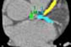
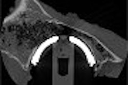
In order to explore the detectability limits of low-dose CT of the thorax and determine the lowest radiation dose possible, Paul and his colleagues developed two phantoms (aptly named “Chester”) that were equipped with simulated nodules of various diameters.
Low-dose CT exams were conducted using a four-row multidetector CT scanner. The researchers conducted three separate phantom studies in which they acquired images at varying kVp (140, 120, 100, and 80 kVp) and mAs (50, 40, 30, 20, and 10 mAs).
Two CT scans (one with nodules and one without) were acquired for each of the three studies. Two radiologists evaluated the ability to detect nodules independently, using contrast-detail and two-alternative forced-choice tests.
“The phantom dose-length product range was 165.12 mGy.cm (140 kVp to 50 mAs) to 8.72 mGy.cm” (80 kVp, 10 mAs), Paul reported. A marked decrease in radiation dose is feasible, he concluded.
In a similar study, researchers also reported that minimum-dose thoracic CT achieves a higher level of sensitivity than chest radiography, specifically in the evaluation of acutely ill patients.
For this pilot study, Dr. Anuradha Rao from the University of Toronto and her team enrolled 100 patients who were admitted to the emergency room with acute dyspnea or chest discomfort.
The researchers performed chest x-ray and a minimum-dose CT protocol at the time of a CT pulmonary angiogram, which was also performed with a reduced-dose technique.
CT scans were performed on four-row and eight-row CT scanners (GE Healthcare, Waukesha, WI). The minimum-dose CT was performed at 120 kVp and 10-30 mAs, which varied according to patient body habitus, Rao reported.
In addition, the group used the CT pulmonary angiogram as a reference standard to examine consolidation, atelectasis, nodules, lymphadenopathy, pleural and pericardial effusions (75 mm) and pulmonary arterial hypertension.
Two staff thoracic radiologists reviewed each study independently. Researchers found that chest x-ray was insensitive in the detection of features for which minimum-dose CT remained sensitive. Comparing the two in terms of lymphadenopathy, the sensitivity was 42% with chest x-ray compared to 100% sensitivity with minimum-dose CT. Pericardial effusions were read with 60% sensitivity on chest x-ray, compared to 100% sensitivity with minimum-dose CT. For pulmonary arterial hypertension, x-ray and CT garnered 80% and 95% sensitivity, respectively.
These features are routinely evaluated in acutely ill patients, Rao said, leading her group to conclude that minimum-dose CT may be a more useful “first-line imaging test above chest x-ray.” However, she cautioned that the findings are part of a pilot study and will require additional data.
Both Rao and Paul said more research is critical, though the refining of low-dose CT techniques is well underway.
By Jerry IngramAuntMinnie.com contributing writer
May 6, 2004
Related Reading
CAD gets role in new ELCAP lung study, April 29, 2004
Mass-based CT calcium scoring leaves room for dose reduction, April 30, 2004
Constant image-noise standard lowers CT dose in pediatric exams, April 21, 2004
Chest x-ray benefits from CAD for spotting lung nodules, March 18, 2004
Lung nodule analysis makes progress, not perfection, March 5, 2004
Copyright © 2004 AuntMinnie.com