VIENNA - Radiologists at the University of Pisa in Italy have found high-resolution MDCT to be a reliable predictor of the degree of vascular infiltration in pancreatic cancer, light years ahead of what single-slice exams once produced, and comparable to US-guided laparoscopy.
"We aimed to evaluate the role of multislice CT in improving our assessment of vascular involvement in pancreatic tumors," said Dr. Davide Caramella in a presentation today at the European Congress of Radiology. "The advantages of MDCT are well known, particularly the high speed of acquisition that allows different studies in different phases of contrast administration."
The ability to visualize tumors in different phases is particularly useful in some hypervascular lesions, for example, which can easily be spotted in the early arterial phase, but not in later stages of contrast distribution, he said.
Predicting the extent of vascular involvement is important for determining the utility of resection.
The study group comprised 59 consecutive patients with suspected pancreatic cancer. Following gastroduodenal distension using three-fourths to one liter of water and pharmacologically induced hypotonia, a high-iodine-concentration contrast material was administered intravenously (110-130 mmol/kg lomeron), and all of the patients underwent thin-section MDCT imaging on a LightSpeed Plus scanner (GE Healthcare, Waukesha, WI).
The group acquired early arterial-phase images and pancreatic-phase images using a slice thickness of 2.5 mm and reconstruction interval of 1.25 mm. Then images were acquired in the venous phase with slice thickness reduced to 1.25 mm, and reconstruction interval of 0.6 mm, he said
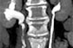
"Image processing was routinely performed, in particular multiplanar reconstructions for highlighting difficult anatomic details, such as in this case, tumor occupying the distal part of the common bile duct," he said. "The MIP (maximum intensity projection) was particularly helpful in depicting the relationship between the tumor and the vascular structures."
The grading of images for vascular involvement was as follows:
- Grade 0: No contact between lesion and vessel
- Grade 1: Focal contiguity between lesion and vessel of less than 50% of the vessel, without affecting the caliber of the vessel
- Grade 2: Focal contiguity of 50% or more, without affecting the caliber of the vessel
- Grade 3: Lesion surrounding the vessel with reduction or obstruction of the lumen
"For our surgical colleagues there are some critical vessels that need special attention from us," he said. "On the arterial side the celiac trunk and the superior mesenteric artery, on the portal side the portal vein and the superior mesenteric vein, so when the tumor arises close to these vessels, it’s particularly important to be very precise in the description."
Of the 59 patients, 37 were considered eligible for surgery, and they underwent pancreatic duodenectomy in 19 cases, distal pancreatectomy in nine cases, and total pancreatectomy in nine. In all there were 28 patients with adenocarcinoma, six with mucinous tumors, and other carcinomas in three cases. Eighteen vessels were classified as zero, six were graded 1, five were graded 2 and three received a grade of 3 for a high probability of vascular infiltration.
There was excellent agreement between MDCT and surgical results, he said. The five cases graded 0 were all correctly identified. In the five cases graded 2, the presence of vascular infiltration was confirmed in four. Of eleven vessels graded 3, 10 showed vascular infiltration and one was a false-positive -- "'fibrotic changes that demonstrated a reduction of the caliber of the vessel that we assumed was due to vascular infiltration, and that was not the case," Caramella said.
Seventeen patients underwent revascularization on these 20 vessels, representing the portal vein in nine cases, the portal mesenteric junction in four, the superior mesenteric vein in five, and the superior mesenteric artery in two, he said. The pathologic analysis on these resected vessels showed that they always confirmed the interoperative findings, in particular the agreement in grade 0 and grade 1, except for the one case that was overestimated. For the assessment of vascular involvement, MDCT had sensitivity of 100%, specificity of 92%, and overall accuracy of 95%, he said.
"'In our experience, multislice CT for the assessment of vascular infiltration performed quite well, much better than we published with same group of surgeons using single-slice CT," he said. "Our present data are very similar to those that we were able to achieve with laparoscopic contact ultrasound. Multislice CT can be an accurate technique (for assessing) pancreatic involvement by pancreatic tumor, and we think that vascular infiltration is very likely only when the neoplasm surrounds the vessel, and this confirms what had already been said in 1997, that a single contiguity of tumor to vessel does not automatically signify vessel invasion."
An audience member inquired as to the use of an MDCT assessment when a surgical team is very agressive. Caramella responded that putting together a database of pathologically proven cases is one way of demonstrating MDCT’s accuracy to surgeons.
By Eric BarnesAuntMinnie.com staff writer
March 6, 2004
Related Reading
Ultrasound of the pancreas: Cystic and non-focal pathology, February 13, 2004
MR, CT detect cystic pancreatic lesions, but malignancy a tough call, February 5, 2004
Mayo group seeks to optimize pancreatic MDCT protocols, October 27, 2003
Copyright © 2004 AuntMinnie.com