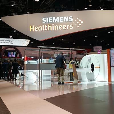
Siemens Healthineers has received approval from the U.S. Food and Drug Administration (FDA) for its high-definition digital breast tomosynthesis (DBT) technology.
High-definition breast tomosynthesis builds on the company's existing DBT platform but offers a tomo sweep of 50° to deliver higher-resolution images, according to the company. It is available as an option on the company's Mammomat Inspiration digital mammography system.
The high-definition tomo mode also incorporates a new version of enhanced multiple parameter iterative reconstruction (EMPIRE), Siemens' data algorithm for mammography. EMPIRE uses iterative reconstruction rather than filtered back projection for improved sensitivity in the detection and diagnosis of breast cancer compared to 2D mammography, according to the company.
While EMPIRE technology has been approved as a 3D-only exam, it also includes Insight 2D and 3D synthetic software-generated visualization of tomosynthesis volumes in both 2D and 3D. Insight 2D allows breast imagers to obtain a 2D image from the EMPIRE 3D image set to provide a navigational support tool to 3D-only exams without subjecting patients to added dose.


![A normal mammogram confirmed by three-year radiologic follow-up illustrates reader-marked regions of interest (ROIs) during (A) unaided (round 1) and (B) artificial intelligence (AI)–assisted (round 2) reading. Each colored dot represents an ROI for recall by a human reader. Readers could mark more than one ROI per case, represented by multiple dots of the same color. During AI-assisted reading, the AI system displayed three visible prompts: two with suspicion of malignancy scores of 35% (left mediolateral oblique [L MLO] and craniocaudal [L CC]) and one with a suspicion of malignancy score of 10% (right craniocaudal [R CC]), shown as polygonal overlays. Without AI, six of 10 readers (60%) marked a false-positive ROI. With AI assistance, this fell to two of 10 (20%). R MLO = right mediolateral oblique.](https://img.auntminnie.com/mindful/smg/workspaces/default/uploads/2026/07/2026-07-14-radiology-mammogram-ai-auto-bias.H0bYO8QlWs.jpg?auto=format%2Ccompress&fit=crop&h=100&q=70&w=100)


![A normal mammogram confirmed by three-year radiologic follow-up illustrates reader-marked regions of interest (ROIs) during (A) unaided (round 1) and (B) artificial intelligence (AI)–assisted (round 2) reading. Each colored dot represents an ROI for recall by a human reader. Readers could mark more than one ROI per case, represented by multiple dots of the same color. During AI-assisted reading, the AI system displayed three visible prompts: two with suspicion of malignancy scores of 35% (left mediolateral oblique [L MLO] and craniocaudal [L CC]) and one with a suspicion of malignancy score of 10% (right craniocaudal [R CC]), shown as polygonal overlays. Without AI, six of 10 readers (60%) marked a false-positive ROI. With AI assistance, this fell to two of 10 (20%). R MLO = right mediolateral oblique.](https://img.auntminnie.com/mindful/smg/workspaces/default/uploads/2026/07/2026-07-14-radiology-mammogram-ai-auto-bias.H0bYO8QlWs.jpg?auto=format%2Ccompress&fit=crop&h=112&q=70&w=112)