Nontraditional education resources are being used more by point-of-care ultrasound (POCUS) educators, a study presented at the 2025 American Institute of Ultrasound in Medicine (AIUM) annual conference found.
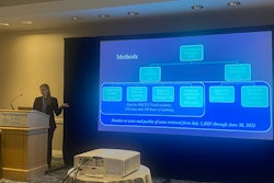
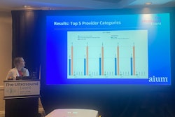
Arthur Broadstock, MD, from the University of Cincinnati in Ohio, presented his team’s results, showing that while POCUS educators still hold in-person workshops in high regard, online media resources are being used more for training medical professionals to use the modality.
"I think the fact that a lot of these resources are free and open-access is appealing to educators, as they can disseminate these resources easily to learners, who have a lower barrier to entry to access this knowledge," Broadstock told AuntMinnie.com. "Ultrasound education is also unique in that much of it depends on review of video clips, which is best served through digital platforms rather than through print materials."
Medical educators are continuing to find ways beyond traditional textbooks to train aspiring professionals. This goes for POCUS education as well, where Broadstock said such resources are abundant. However, he pointed out that the extent to which educators use these nontraditional resources is not well known.
 Arthur Broadstock, MD, presents research at AIUM 2025 showing how point-of-care ultrasound (POCUS) eduactors are using more nontraditional learning resources in their curricula.
Broadstock and colleagues conducted a survey study to better understand what materials POCUS educators prefer to use for training.
Final analysis included 62 completed surveys by educators. The team used a six-point Likert-type scale to gauge the importance of different educational materials as rated by survey respondents.
Educators listed in-person workshops and online media resources as highly important while textbooks were of least importance, Broadstock reported.
| Importance of POCUS educational resources as rated by educators | |
|---|---|
| Resources | Likert score (out of 6) |
| In-person workshops | 5.1 |
| Online media | 4.8 |
| Journal articles | 3.4 |
| eBooks | 2.9 |
| Podcasts | 2.8 |
| Textbooks | 2.1 |
The researchers also examined specific resources that educators use in their curricula. For traditional resources, the following were the most commonly used among educators: Point of Care Ultrasound in Critical Care (100%), Emergency Ultrasound Made Easy (98.4%), and Pediatric Emergency and Critical Care Ultrasound (88.9%).
For nontraditional resources, this trend included the following: Core Ultrasound/5 Minute Sono/Ultrasound of the Week (87.3%), Ultrasound GEL Podcast (65.1%), ACEP Sonoguide (41.9%), and SAEM Academy of Emergency Ultrasound Narrated Lecture Series (34.9%). These findings highlight that there’s a strong reliance on online open-access resources alongside a few key texts, Broadstock said.
He added that in-person workshops may still be held in high regard by educators due to the hands-on nature of ultrasound.
"Performing an ultrasound exam is inherently a psychomotor skill, so in-person training is necessary to build the image acquisition component of POCUS and I think educators understand that, and it was reflected in the rankings," Broadstock told AuntMinnie.com.
He concluded that future research should explore resource use by POCUS learners and assess the quality of digital resources.
“As POCUS education evolves, a blended learning approach combining hands-on experience with high-quality digital materials may provide the most effective training,” he added.