ORLANDO -- Point-of-care ultrasound (POCUS) residency tracks lead to marked improvement in exam performance by family medicine specialists, according to findings presented March 31 at the 2025 American Institute of Ultrasound in Medicine (AIUM) annual conference.
In her talk, Laurel Gutierrez, MD, from the University of Michigan in Ann Arbor, discussed her team’s research on how first-year residents significantly performed better on POCUS exams than non-track residents at their institution.
“The ultimate goal is that we prepare family medicine residents to be able to use POCUS in their practice,” Gutierrez said.
POCUS continues to make its way into training curricula for medical students and residents. In 2021, nearly one-third of family medicine residencies reported having an established POCUS curriculum.
However, the lack of POCUS imaging standards may limit the effectiveness of these efforts. Gutierrez said an important part of quality improvement that should be in all POCUS programs is performing quality analysis of POCUS scans, with blinded review based on established criteria being the standard.
She further noted that there is little data available on scanning volume or the types of scans performed within family medicine residencies.
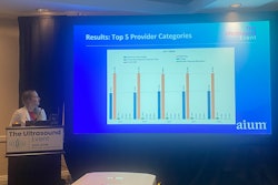
Laurel Gutierrez, MD, at AIUM 2025 presents results on how track residents for a POCUS program saw their skills with the modality improved more significantly than that of nontrack residents in family medicine.
Gutierrez and colleagues developed their POCUS residency track for family medicine residents to improve their scan quality performance.
The pilot program included three track residents who underwent the following during their track time in 2021 and 2022: seven simulation sessions, elective POCUS scanning time, quarterly POCUS training, and biannual simulation sessions. The program developers compared the results of the track residents to those of nontrack residents (n = 36) who only underwent quarterly training and biannual simulation sessions.
Gutierrez and fellow researchers also compared the quality of images from the first six months of the track to images from the last six months.
Track residents performed a total of 222 scans in the year-long program compared with 63 for the nontrack residents. The analysis included eight scan types, the most common of which performed by track residents included the following: musculoskeletal (n = 75), soft tissue (n = 68), and renal and/or bladder (n = 17).
Track residents showed significant improvement in all scan types. These included soft tissue (p < 0.01), lung (p < 0.001), shoulder (p < 0.01), knee (p < 0.01), cardiovascular (p = 0.01), abdominal aortic aneurysm (p < 0.001), gallbladder (p = 0.01), and renal (p < 0.001).
Nontrack residents meanwhile significantly improved in only soft tissue, shoulder, and abdominal aortic aneurysm scans (p < 0.05 for all).
Gutierrez said the results show that some POCUS scanning types require dedicated practice and more extensive training. She added that participation in didactics may have contributed to significant improvement in some areas for nontrack residents.
She called for future efforts to define acceptable quality for POCUS images and increase the number of scans for track residents.
“We do need to continue to assess learning outcomes for track residents … and also address barriers for POCUS use,” Gutierrez said.


![Examples of ultrasound findings and techniques. (A) Images in a 39-year-old male patient with a mass in the left thigh. The mass is heterogeneous on the B-mode US image (compared with the patient in D) and showed increased microvascularity (superb microvascular imaging [SMI]) and shear-wave elastography (SWE) values. Undifferentiated pleomorphic sarcoma was diagnosed at biopsy (with pleomorphic rhabdomyosarcoma in surgical specimen). (B) Images in an 18-year-old male patient with a mass in the left leg. The mass is hypoechoic on the B-mode image, with no other findings suggestive of malignancy. The lesion is in contact with the cortex of the tibia, which is slightly irregular. CT revealed a doubtful anteromedial tibial erosion. The microvascular study demonstrated high vascularization, suggestive of malignancy. Periosteal Ewing sarcoma was diagnosed with both histologic and immunohistochemical confirmation. (C) Images in a 69-year-old female patient with a lump growing on the outside of the left leg. Multiple SWE examinations were performed (please note the high values obtained in the measurements, whereas the color map highlights the stiffness relative to adjacent tissues). SMI showed areas of increased vascularization to target for sampling. Undifferentiated spindle cell sarcoma was diagnosed at biopsy, with residual leiomyosarcoma in the surgical specimen after neoadjuvant therapy. (D) Images in a 56-year-old female patient with a mass in the right thigh. The mass is heterogeneous at both B-mode ultrasound (similar to patient A) and MRI (coronal T2-weighted spectral attenuated inversion recovery [SPAIR]; T1-weighted pre-contrast and postcontrast imaging), which even shows uptake after the administration of paramagnetic contrast material, which is traditionally suggestive of malignancy. Low values at SMI and elastography are suggestive of benignity. Spindle cell lipoma was diagnosed at biopsy, with atypical spindle cell lipomatous tumor in the surgical specimen.](https://img.auntminnie.com/mindful/smg/workspaces/default/uploads/2026/08/images-radiol250278fig2.APCFLSvX6p.jpg?auto=format%2Ccompress&fit=crop&h=100&q=70&w=100)


![Examples of ultrasound findings and techniques. (A) Images in a 39-year-old male patient with a mass in the left thigh. The mass is heterogeneous on the B-mode US image (compared with the patient in D) and showed increased microvascularity (superb microvascular imaging [SMI]) and shear-wave elastography (SWE) values. Undifferentiated pleomorphic sarcoma was diagnosed at biopsy (with pleomorphic rhabdomyosarcoma in surgical specimen). (B) Images in an 18-year-old male patient with a mass in the left leg. The mass is hypoechoic on the B-mode image, with no other findings suggestive of malignancy. The lesion is in contact with the cortex of the tibia, which is slightly irregular. CT revealed a doubtful anteromedial tibial erosion. The microvascular study demonstrated high vascularization, suggestive of malignancy. Periosteal Ewing sarcoma was diagnosed with both histologic and immunohistochemical confirmation. (C) Images in a 69-year-old female patient with a lump growing on the outside of the left leg. Multiple SWE examinations were performed (please note the high values obtained in the measurements, whereas the color map highlights the stiffness relative to adjacent tissues). SMI showed areas of increased vascularization to target for sampling. Undifferentiated spindle cell sarcoma was diagnosed at biopsy, with residual leiomyosarcoma in the surgical specimen after neoadjuvant therapy. (D) Images in a 56-year-old female patient with a mass in the right thigh. The mass is heterogeneous at both B-mode ultrasound (similar to patient A) and MRI (coronal T2-weighted spectral attenuated inversion recovery [SPAIR]; T1-weighted pre-contrast and postcontrast imaging), which even shows uptake after the administration of paramagnetic contrast material, which is traditionally suggestive of malignancy. Low values at SMI and elastography are suggestive of benignity. Spindle cell lipoma was diagnosed at biopsy, with atypical spindle cell lipomatous tumor in the surgical specimen.](https://img.auntminnie.com/mindful/smg/workspaces/default/uploads/2026/08/images-radiol250278fig2.APCFLSvX6p.jpg?auto=format%2Ccompress&fit=crop&h=112&q=70&w=112)