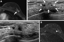
Ultrasound can play a significant role in cancer detection for women with dense breast tissue where access to supplemental screening with MRI and/or contrast mammography is limited, researchers have reported.
In a study conducted by Paula Gordon, MD, and Linda Warren, MD, both of the University of British Columbia in Vancouver, and colleague Jean Seely, MD, of the University of Ottawa, the modality showed a high incremental cancer detection rate (ICDR) in women with both heterogeneously dense and extremely dense tissue who underwent annual 2D mammography screening. The findings were published February 21 in the Canadian Association of Radiologists Journal.
Breast ultrasound is one of a variety of tools proposed for supplemental screening of women with dense breasts, but it is not widely available in Canada, Gordon and colleagues noted. They investigated the efficacy of supplemental screening breast ultrasound in women with heterogeneously dense and extremely dense tissue, conducting a study that assessed ultrasound-guided breast biopsies prompted by screening breast ultrasound performed from August 2021 to December 2022. The team assessed risk factors, disease stage, ICDR, biopsy rates, and positive predictive values for biopsy (PPV3). Of 5,257 women screened, 247 underwent biopsies, and of these, 32 were diagnosed with breast cancer (27 invasive and 5 ductal carcinoma in situ).
Gordon's group reported a positive predictive value for biopsy of 13% and an incidental cancer detection rate of 6.1 out of 1,000 women screened with breast ultrasound. It also found the following:
- 15.6% of DCIS were stage 0; 56.3% were stage 1A; and 6.3% were stage 1B.
- 3.1% of ductal/lobular cancers were stage 3B; 16.6% of lobular cancers were stage 1A, 1B, or 2B.
- 3.1% of adenoid cystic carcinomas were stage 2A.
- Three cancers were found on incident screens and 29 on prevalent screens; of these, 84.4% were in women with heterogeneously dense tissue and 62.5% were found in women with no personal or first-degree family history of breast cancer.
"This essential lifesaving examination should be available for women with dense breasts to give them equity in the early detection of breast cancer, and fee codes should be created to incentivize radiologists to offer the service," the team concluded.
The complete study can be found here.



![Examples of ultrasound findings and techniques. (A) Images in a 39-year-old male patient with a mass in the left thigh. The mass is heterogeneous on the B-mode US image (compared with the patient in D) and showed increased microvascularity (superb microvascular imaging [SMI]) and shear-wave elastography (SWE) values. Undifferentiated pleomorphic sarcoma was diagnosed at biopsy (with pleomorphic rhabdomyosarcoma in surgical specimen). (B) Images in an 18-year-old male patient with a mass in the left leg. The mass is hypoechoic on the B-mode image, with no other findings suggestive of malignancy. The lesion is in contact with the cortex of the tibia, which is slightly irregular. CT revealed a doubtful anteromedial tibial erosion. The microvascular study demonstrated high vascularization, suggestive of malignancy. Periosteal Ewing sarcoma was diagnosed with both histologic and immunohistochemical confirmation. (C) Images in a 69-year-old female patient with a lump growing on the outside of the left leg. Multiple SWE examinations were performed (please note the high values obtained in the measurements, whereas the color map highlights the stiffness relative to adjacent tissues). SMI showed areas of increased vascularization to target for sampling. Undifferentiated spindle cell sarcoma was diagnosed at biopsy, with residual leiomyosarcoma in the surgical specimen after neoadjuvant therapy. (D) Images in a 56-year-old female patient with a mass in the right thigh. The mass is heterogeneous at both B-mode ultrasound (similar to patient A) and MRI (coronal T2-weighted spectral attenuated inversion recovery [SPAIR]; T1-weighted pre-contrast and postcontrast imaging), which even shows uptake after the administration of paramagnetic contrast material, which is traditionally suggestive of malignancy. Low values at SMI and elastography are suggestive of benignity. Spindle cell lipoma was diagnosed at biopsy, with atypical spindle cell lipomatous tumor in the surgical specimen.](https://img.auntminnie.com/mindful/smg/workspaces/default/uploads/2026/08/images-radiol250278fig2.APCFLSvX6p.jpg?auto=format%2Ccompress&fit=crop&h=100&q=70&w=100)


![Examples of ultrasound findings and techniques. (A) Images in a 39-year-old male patient with a mass in the left thigh. The mass is heterogeneous on the B-mode US image (compared with the patient in D) and showed increased microvascularity (superb microvascular imaging [SMI]) and shear-wave elastography (SWE) values. Undifferentiated pleomorphic sarcoma was diagnosed at biopsy (with pleomorphic rhabdomyosarcoma in surgical specimen). (B) Images in an 18-year-old male patient with a mass in the left leg. The mass is hypoechoic on the B-mode image, with no other findings suggestive of malignancy. The lesion is in contact with the cortex of the tibia, which is slightly irregular. CT revealed a doubtful anteromedial tibial erosion. The microvascular study demonstrated high vascularization, suggestive of malignancy. Periosteal Ewing sarcoma was diagnosed with both histologic and immunohistochemical confirmation. (C) Images in a 69-year-old female patient with a lump growing on the outside of the left leg. Multiple SWE examinations were performed (please note the high values obtained in the measurements, whereas the color map highlights the stiffness relative to adjacent tissues). SMI showed areas of increased vascularization to target for sampling. Undifferentiated spindle cell sarcoma was diagnosed at biopsy, with residual leiomyosarcoma in the surgical specimen after neoadjuvant therapy. (D) Images in a 56-year-old female patient with a mass in the right thigh. The mass is heterogeneous at both B-mode ultrasound (similar to patient A) and MRI (coronal T2-weighted spectral attenuated inversion recovery [SPAIR]; T1-weighted pre-contrast and postcontrast imaging), which even shows uptake after the administration of paramagnetic contrast material, which is traditionally suggestive of malignancy. Low values at SMI and elastography are suggestive of benignity. Spindle cell lipoma was diagnosed at biopsy, with atypical spindle cell lipomatous tumor in the surgical specimen.](https://img.auntminnie.com/mindful/smg/workspaces/default/uploads/2026/08/images-radiol250278fig2.APCFLSvX6p.jpg?auto=format%2Ccompress&fit=crop&h=112&q=70&w=112)