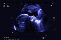
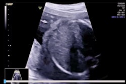
Point-of-care ultrasound (POCUS) can be a useful tool for diagnosing "retained products of conception" in women, suggest findings published January 13 in the American Journal of Emergency Medicine.
A team led by Zachary Boivin, MD, from Yale University in New Haven, CT, found that POCUS can accurately diagnose this tissue in patients being managed surgically or medically.
“Emergency physicians should use POCUS as a first-line imaging modality in patients with history concerning for [retained products of conception] to rule in a time-sensitive diagnosis,” the Boivin team wrote.
"Retained products of conception" refers to residual intrauterine tissue in women that can occur after a recent birth, pregnancy termination, or miscarriage. These occur in a range of 1% to 6% of all pregnancies. However, incidence increases with second-trimester deliveries and pregnancy terminations. Once diagnosed, women are treated via dilation and curettage, the drug misoprostol, or ongoing follow-up.
Ultrasound is the go-to method for diagnosing tissue retained as the result of conception. However, the researchers noted that there are varied diagnostic criteria for using ultrasound in this area and little consensus among previous studies. They added that there is a lack of research that systematically examines how emergency physicians diagnose retained products of conception via POCUS.
To address the knowledge gap, Boivin and colleagues investigated the test characteristics of POCUS in detecting this particular tissue and evaluated how cases were managed in the emergency department.
The study included data collected between 2017 and 2023 from 645 women. For ground truth testing, 512 of the women underwent standard nonportable ultrasound while the remaining 133 underwent non-imaging-related ob/gyn testing. Through these, ultrasound helped find 42 cases of retained products of conception while OB/GYN testing led to 20 confirmed cases. At a total of 62 women, retained products of conception had an incidence rate of 9.6% in the entire study cohort.
The team reported 70 total POCUS exams that identified retained products of conception, with 17 indeterminate exams.
Of the total women, 265 had a history that led to concerns about retained products of conception. In these women, POCUS achieved a sensitivity of 79% and specificity of 93.8% compared to the gold standard diagnosis.
Eight of the POCUS exams in this group were deemed indeterminate. The team reported that if all these indeterminate exams were considered positive, the sensitivity would be 80.7% and specificity would be 90.4 %.
Finally, of the 62 total patients diagnosed via chart review, 21 were admitted, 26 had a surgical procedure, 26 were managed medically, and 10 were surveilled.
Despite the promising results, the study authors cautioned that POCUS use in early pregnancy could lead to false-positive findings, given the variable appearance of the endometrium during this time. They added that numerous diagnoses can mimic retained products of conception on blinded POCUS review.
The team reported 22 false positives on blinded POCUS review, of which over half were from potentially viable pregnancies when reviewing the ground truth tests.
“Further prospective studies are needed to confirm our findings and determine if POCUS can reduce patient length of stay and time to management,” the authors concluded.
The full study can be accessed here.