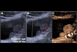
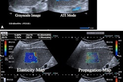
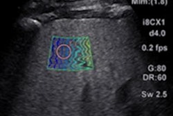
Endoscopic ultrasound shear wave elastography (SWE) can detect clinically significant portal hypertension (CSPH) in the spleen, suggest findings published on September 7 in iGIE.
Researchers led by Vinay Chandrasekhara, MD, from the Mayo Clinic in Rochester, MN, reported that patients with CSPH had significantly higher spleen stiffness compared with those without hypertension as observed on SWE.
"SWE is a promising technology that can readily be incorporated into standard endoscopic ultrasound examination for assessment portal hypertension," Chandrasekhara and colleagues wrote.
CSPH is a condition that causes spleen deterioration and potentially fatal complications, especially in the presence of advanced liver disease, such as cirrhosis. This makes the early detection of CSPH important to inform patient management and monitoring.
While conventional testing is invasive and requires vascular access, researchers have explored the potential of noninvasive testing methods such as ultrasound and MRI. The researchers highlighted that recent upgrades of endoscopic ultrasound processors now allow for liver and spleen stiffness measuring via SWE.
The Chandrasekhara team studied SWE's performance in measuring spleen stiffness to detect CSPH and defined test characteristics for future research.
The final analysis included data collected between 2020 and 2023 from 142 patients, all of whom underwent SWE. Of the patients, 13 had CSPH while the remaining 129 did not. The researchers noted no significant differences among the patients when it came to age, sex, and body mass index. Also, 84 of the patients were sedated with general anesthesia and 58 underwent monitored anesthesia care.
Eight of the 13 patients with CSPH had liver disease tied to either alcohol or metabolic dysfunction, while one other patient had noncirrhotic portal hypertension.
Patients with CSPH had higher average liver and spleen stiffness compared with patients without CSPH. Additionally, the team found no significant differences between shear wave velocity and reliability index between the two patient cohorts.
| SWE performance in spleen, liver stiffness measurements | |||
|---|---|---|---|
| Measure | Non-CSPH | CSPH | p-value |
| Average FIB4 score | 1.7 | 4.7 | < 0.001 |
| Average liver stiffness (in kPA) | 15.9 | 27 | < 0.001 |
| Average spleen stiffness | |||
| Elastic modulus (average kPA) | 29.1 | 37.6 | 0.003 |
| Shear wave velocity (m/s) | 3 | 3.3 | 0.36 |
| Reliability index | 85.1% | 74% | 0.12 |
Also, patients on average underwent a median of 12 measurements and the area under the receiver operating curve was 0.74. The test cutoff that maximized performance characteristics was 37.6 kPa with a 69.2% sensitivity and 79.8% specificity, the researchers said.
Finally, on univariate linear regression, the team observed that spleen stiffness was significantly correlated with liver stiffness measured obtained through SWE (ß = 0.21 kPa, p = 0.001) as well as magnetic resonance elastography (ß = 0.08 kPa, p = 0.003).
The study authors noted that elastography measurements can provide key information on the status of a patient's liver disease during endoscopy. It also adds "minimal" risk and doesn't require specialized skillsets beyond endoscopic ultrasound.
"The optimal use of endoscopic ultrasound SWE technology would detect CSPH in patients prior to exhibiting clinical/imaging findings or in those with conflicting results," they wrote.
The authors called for prospective studies to validate and optimize diagnostic performance while also determining how spleen stiffness correlates to decompensating events like variceal bleeding.
The full study can be found here.