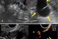
Ultrasound shows that isthmic contractions are common in second-trimester pregnancies and can affect cervical measurements, a Spanish study published July 20 in the American Journal of Obstetrics and Gynecology found.
Researchers led by Alba Farras, MD, PhD, from Vall d'Hebron University Hospital in Barcelona reported ultrasound findings that showed isthmic contractions occur in close to half of pregnant women after bladder voiding, and that it takes about 20 minutes for complete relaxation of the isthmus after a contraction.
"We recommend performing cervical assessment at least 20 minutes after bladder voiding to reduce the risk of bias in cervical length measurement and to avoid false images of placenta previa," Farras and colleagues wrote.
While asymptomatic contractions in the uterine isthmus -- also known as isthmic contractions --are a frequent physiological occurrence in pregnant women, the researchers noted that there is limited data on their prevalence and characteristics. Previous studies have used static ultrasound images, which may not sufficiently capture the nature of these contractions, according to the authors, so they used real-time ultrasound to investigate with an end goal of developing a new way to assess the contractions' presence and intensity.
The study included data from 30-second trimester singleton pregnancies; the women were assessed for isthmic contractions after voiding their bladders. The team found that the contractions occurred in 43% of the women and that the median time to complete isthmus relaxation was 19.7 minutes.
Farras and colleagues also found that relaxation tended to start with a decrease in muscular tone in the isthmus, creating a funnel-like image. This image disappeared as the isthmus continued to relax and uterine wall thickness decreased.
More research is needed to understand the role of isthmic contractions in pregnancy and birth, the authors urged. They recommended the following steps for assessing the presence of these contractions:
- Observe the cervix for at least 3 minutes. If the maximum isthmic length observed during that period is more than 18 mm, clinicians can assume that the patient is experiencing an isthmic contraction.
- If the patient is experiencing an isthmic contraction, wait at least 20 minutes and reassess the cervix.
- For universal cervical length screening performed in the second trimester of pregnancy, have the patient void their bladder, then scan the fetus, and perform the cervical length screening 20 to 30 minutes after voiding.
- Do not wait more than 30 minutes, because bladder filling can falsely exaggerate cervical length.
- Check for signs of isthmic contraction before diagnosing placenta previa.
- If the isthmic length is over 18 mm or subjective signs of contraction are seen, repeat the ultrasound examination 20 minutes later.
The entire study can be found here.