A focused ultrasound treatment that uses microbubbles to amplify ultrasound's effects on the brain's blood vessels may be able to help mitigate brain diseases, according to research published May 15 in Proceedings of the National Academy of Sciences.
Researchers from Washington University in St. Louis led by Dezhuang Ye, PhD, described how their method creates a pumping effect to help remove accumulating brain waste that may cause brain pathologies.
"Findings from this study suggest that ultrasound can be utilized as a noninvasive and nonpharmacological approach to mitigate brain diseases caused by impaired glymphatic function," said corresponding author Dr. Hong Chen from the university.
The brain clears waste such as dead blood cells and excess proteins through the glymphatic system. Impairment to this system may place people more at risk for brain disease pathologies such as stroke, neurodegenerative disease, and traumatic brain injuries.
The researchers believed that ultrasound waves could help with this by noninvasively stimulating mechanical effects on the brain. By using focused ultrasound, which concentrates this energy onto regions of interest, they hypothesized that this method combined with microbubbles from contrast injection could do so with great precision. The authors had previously found that the combined approach improved the penetration and accumulation of intranasally administered drugs, which reach the brain through the olfactory and trigeminal nerve pathways.
The team tested the technique using mouse models, administering a fluorescently labeled albumin tracer through the nasal canal. Then, the researchers intravenously injected microbubbles and administered focused ultrasound waves aimed deep in the brain at the thalamus. They compared results to control groups, which included tracer administration with focused ultrasound but without microbubbles and tracer administration without either.
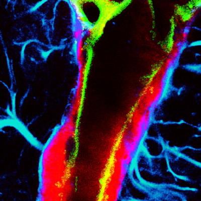
 This microscopic image reveals the enhanced glymphatic transport of an intranasally delivered tracer (red), achieved using ultrasound combined with microbubbles. Image courtesy of the Chen Lab, Washington University in St. Louis.
Through 3D imaging of the treated side of the brain, they found that the combined method improved the transport of the tracer through the blood vessels. They also found evidence of focused ultrasound-enhanced penetration of the tracer from the perivascular space into the interstitial space, primarily along arterioles. The control groups meanwhile showed lower tracer accumulation.
The researchers also used the combined method after injecting the tracer directly to the cerebral spinal fluid. While this method is invasive, the team did so to further validate its results. It found that focused ultrasound with microbubbles improved tracer transport along vessels at the targeted brain site by "about two- to threefold compared with the nontargeted side."
For the arterioles, the researchers found that the average albumin fluorescence intensity in the treated brain region improved by 8.75-fold in the intranasal administration group and 3.49-fold in the ICM group when compared to the nontreated side (p < 0.05 and p < 0.01, respectively). They also noted smaller-scale improvements in the capillaries and venules.
"Regardless of whether tracers were delivered via intranasal or intracisterna magna route, focused ultrasound with microbubbles consistently improved glymphatic transport," the study authors wrote.
The authors suggested that these results open opportunities for ultrasound combined with microbubbles to improve glymphatic transport, as well as hold promise in mitigating neurodegenerative diseases like Alzheimer's and Parkinson's by enhancing waste clearance in the brain.
Chen told AuntMinnie.com that the team plans to use in vivo dynamic imaging tools to reveal the dynamics of agent transport by the ultrasound-microbubble method in the glymphatic system.
"We also plan to determine the impact of different treatment parameters on glymphatic transport and establish the efficacy profiles of focused ultrasound with microbubbles in glymphatic manipulation," she said.