Thursday, December 1 | 8:00 a.m.-9:00 a.m. | R1-SSMK10-1 | E451A
In this presentation, research will highlight the safety and efficacy of MR-guided high-intensity focused ultrasound (MR-HIFU) when it comes to treating osteoid osteoma close to sensitive body structures.Dr. Valerio D'Agostino from the University of Naples Federico II in Naples will talk about his team's research, which analyzed this imaging method in a total of 36 patients. Although the standard for this ultrasound method is for use in a distance greater than 1 cm between the target lesion and sensitive structures, the team wanted to test it in closer proximity to these structures.
Goals of this study included analyzing complications involving sensitive structures, assessing treatment response, and looking at the impact of the proximity of sensitive structures on treatment efficacy.
The researchers found no documented adverse events over grade 2. The average pain score at baseline, measured via the visual analog scale, was 4. This was followed by complete pain relief at clinical follow-up 12 to 24 months for all but three patients. For these three patients, one was re-treated with MR-guided high-intensity focused ultrasound, while the other two were treated with CT-guided radiofrequency ablation.
The researchers also found that location in proximity of sensitive structures did not affect the efficacy of treatments (p < 0.01).
See what else the team found at D'Agostino's presentation.




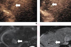



![Examples of ultrasound findings and techniques. (A) Images in a 39-year-old male patient with a mass in the left thigh. The mass is heterogeneous on the B-mode US image (compared with the patient in D) and showed increased microvascularity (superb microvascular imaging [SMI]) and shear-wave elastography (SWE) values. Undifferentiated pleomorphic sarcoma was diagnosed at biopsy (with pleomorphic rhabdomyosarcoma in surgical specimen). (B) Images in an 18-year-old male patient with a mass in the left leg. The mass is hypoechoic on the B-mode image, with no other findings suggestive of malignancy. The lesion is in contact with the cortex of the tibia, which is slightly irregular. CT revealed a doubtful anteromedial tibial erosion. The microvascular study demonstrated high vascularization, suggestive of malignancy. Periosteal Ewing sarcoma was diagnosed with both histologic and immunohistochemical confirmation. (C) Images in a 69-year-old female patient with a lump growing on the outside of the left leg. Multiple SWE examinations were performed (please note the high values obtained in the measurements, whereas the color map highlights the stiffness relative to adjacent tissues). SMI showed areas of increased vascularization to target for sampling. Undifferentiated spindle cell sarcoma was diagnosed at biopsy, with residual leiomyosarcoma in the surgical specimen after neoadjuvant therapy. (D) Images in a 56-year-old female patient with a mass in the right thigh. The mass is heterogeneous at both B-mode ultrasound (similar to patient A) and MRI (coronal T2-weighted spectral attenuated inversion recovery [SPAIR]; T1-weighted pre-contrast and postcontrast imaging), which even shows uptake after the administration of paramagnetic contrast material, which is traditionally suggestive of malignancy. Low values at SMI and elastography are suggestive of benignity. Spindle cell lipoma was diagnosed at biopsy, with atypical spindle cell lipomatous tumor in the surgical specimen.](https://img.auntminnie.com/mindful/smg/workspaces/default/uploads/2026/08/images-radiol250278fig2.APCFLSvX6p.jpg?auto=format%2Ccompress&fit=crop&h=112&q=70&w=112)


