Dear Ultrasound Insider,
The dog days of summer are among us, so maybe it's a good time to stay in the lab or clinic while the outside world deals with the extreme heat. Perhaps the extra time indoors can spur innovation. In the meantime, ultrasound has witnessed some advancements that can be seen in the clinic or even in your own kitchen.
A team from Nottingham City Hospital in England used common kitchen ingredients such as corn flour, gelatin, and tofu to create an ultrasound phantom that mimics breast tissue. How did trainees respond to making their own phantoms with these ingredients? Find out in this edition's Insider Exclusive.
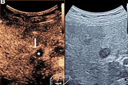
In other news, contrast-enhanced ultrasound (CEUS) continues to gain traction with one of the largest prospective studies to date. A Chinese group found that CEUS-guided biopsy is superior to biopsies guided by conventional ultrasound when it comes to focal liver lesions.
Also, investigators found that a nomogram predictive model using CEUS can help predict prostate cancer in men. The model, which also uses age and the Prostate Image and Reporting Data System (PI-RADS), showed a higher area under the curve than control models.
In sportier news, point-of-care ultrasound has been up to serve at the US Open tennis tournament for several years now. But researchers recently highlighted their experience in using the technology at the annual tournament, writing that it helps streamline medical care for athletes.
Plus, a Spanish team developed an artificial intelligence (AI) model trained with ultrasound images that the group said can help differentiate between malignant lymph nodes and benign nodes that react as a side effect of COVID-19 vaccination.
Plus, check out the following recent ultrasound stories:
- A research team from China recently developed an AI model that can create x-ray-like images from ultrasound data, helping visualize adolescent idiopathic scoliosis.
- A Russian cruise missile attack in July killed at least 24 civilians and wounded dozens more, including radiology staff members at a clinic in Vinnytsia, Ukraine. The clinic housed one 1.5-tesla MRI unit, three ultrasound machines, and five rooms for specialty consultations and was also destroyed in the attack.
- A Canadian group found that microultrasound has detection rates for prostate cancer that are comparable to multiparametric MRI in men who have not yet been biopsied.
- Researchers who presented at the recent ECR shed more light on lymphadenopathy caused by the COVID-19 vaccines, with ultrasound scans helping show this reaction.
Find more articles like these by regularly visiting our Ultrasound Community!



![Examples of ultrasound findings and techniques. (A) Images in a 39-year-old male patient with a mass in the left thigh. The mass is heterogeneous on the B-mode US image (compared with the patient in D) and showed increased microvascularity (superb microvascular imaging [SMI]) and shear-wave elastography (SWE) values. Undifferentiated pleomorphic sarcoma was diagnosed at biopsy (with pleomorphic rhabdomyosarcoma in surgical specimen). (B) Images in an 18-year-old male patient with a mass in the left leg. The mass is hypoechoic on the B-mode image, with no other findings suggestive of malignancy. The lesion is in contact with the cortex of the tibia, which is slightly irregular. CT revealed a doubtful anteromedial tibial erosion. The microvascular study demonstrated high vascularization, suggestive of malignancy. Periosteal Ewing sarcoma was diagnosed with both histologic and immunohistochemical confirmation. (C) Images in a 69-year-old female patient with a lump growing on the outside of the left leg. Multiple SWE examinations were performed (please note the high values obtained in the measurements, whereas the color map highlights the stiffness relative to adjacent tissues). SMI showed areas of increased vascularization to target for sampling. Undifferentiated spindle cell sarcoma was diagnosed at biopsy, with residual leiomyosarcoma in the surgical specimen after neoadjuvant therapy. (D) Images in a 56-year-old female patient with a mass in the right thigh. The mass is heterogeneous at both B-mode ultrasound (similar to patient A) and MRI (coronal T2-weighted spectral attenuated inversion recovery [SPAIR]; T1-weighted pre-contrast and postcontrast imaging), which even shows uptake after the administration of paramagnetic contrast material, which is traditionally suggestive of malignancy. Low values at SMI and elastography are suggestive of benignity. Spindle cell lipoma was diagnosed at biopsy, with atypical spindle cell lipomatous tumor in the surgical specimen.](https://img.auntminnie.com/mindful/smg/workspaces/default/uploads/2026/08/images-radiol250278fig2.APCFLSvX6p.jpg?auto=format%2Ccompress&fit=crop&h=100&q=70&w=100)


![Examples of ultrasound findings and techniques. (A) Images in a 39-year-old male patient with a mass in the left thigh. The mass is heterogeneous on the B-mode US image (compared with the patient in D) and showed increased microvascularity (superb microvascular imaging [SMI]) and shear-wave elastography (SWE) values. Undifferentiated pleomorphic sarcoma was diagnosed at biopsy (with pleomorphic rhabdomyosarcoma in surgical specimen). (B) Images in an 18-year-old male patient with a mass in the left leg. The mass is hypoechoic on the B-mode image, with no other findings suggestive of malignancy. The lesion is in contact with the cortex of the tibia, which is slightly irregular. CT revealed a doubtful anteromedial tibial erosion. The microvascular study demonstrated high vascularization, suggestive of malignancy. Periosteal Ewing sarcoma was diagnosed with both histologic and immunohistochemical confirmation. (C) Images in a 69-year-old female patient with a lump growing on the outside of the left leg. Multiple SWE examinations were performed (please note the high values obtained in the measurements, whereas the color map highlights the stiffness relative to adjacent tissues). SMI showed areas of increased vascularization to target for sampling. Undifferentiated spindle cell sarcoma was diagnosed at biopsy, with residual leiomyosarcoma in the surgical specimen after neoadjuvant therapy. (D) Images in a 56-year-old female patient with a mass in the right thigh. The mass is heterogeneous at both B-mode ultrasound (similar to patient A) and MRI (coronal T2-weighted spectral attenuated inversion recovery [SPAIR]; T1-weighted pre-contrast and postcontrast imaging), which even shows uptake after the administration of paramagnetic contrast material, which is traditionally suggestive of malignancy. Low values at SMI and elastography are suggestive of benignity. Spindle cell lipoma was diagnosed at biopsy, with atypical spindle cell lipomatous tumor in the surgical specimen.](https://img.auntminnie.com/mindful/smg/workspaces/default/uploads/2026/08/images-radiol250278fig2.APCFLSvX6p.jpg?auto=format%2Ccompress&fit=crop&h=112&q=70&w=112)