Dear Ultrasound Insider,
Newer uses for ultrasound are getting their time in the spotlight as researchers continue to explore novel uses for elastography, contrast enhancements, and ... ultrasound gel for hair?
Before we get to that last part, the American College of Radiology in 2018 created the Ovarian-Adnexal Reporting and Data System (O-RADS) ultrasound risk stratification system to help determine the risk of adnexal lesions. Researchers across the U.S. studied the diagnostic performance of this system on women presenting at radiology departments in the country. Find out how well it did in this edition's Insider Exclusive.
Now for that hair gel part. An Italian research team found that applying ultrasound gel to a patient's hair can contain tresses and prevent hair from invading a surgical site. Read more to learn how this could be useful for patients undergoing craniofacial or maxillofacial surgery.
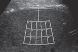
In other news, Chinese researchers found that a method of shear-wave ultrasound imaging called sound touch elastography can help identify breast malignancies by measuring tissue stiffness around lesions. They wrote that this method achieved high marks by reflecting collagen fiber content in malignant tissue.
Also, contrast-enhanced ultrasound (CEUS) can characterize soft-tissue vascular abnormalities more accurately than color Doppler, according to a recent study. The study authors wrote that CEUS also increases diagnostic confidence and is safe for patients, which could reduce the need for CT or MRI scans.
Plus, check out the following recent ultrasound stories:
- Researchers from the Cincinnati Children's Hospital Medical Center found that ultrasound shear-wave elastography can correlate well with MR elastography for measuring liver stiffness in children and young adults with known or suspected liver disease.
- Presenters at the American Roentgen Ray Society (ARRS) meeting in New Orleans showed that classifying lesions found on ultrasound through the Thyroid Imaging Reporting and Data System (TI-RADS) is good, but improvements can be made.
- Another presentation at ARRS showed that ultrasound of the chest wall performs well in follow-up imaging of breast cancer patients after mastectomy.
- Over half of women with axillary lymphadenopathy after receiving the COVID-19 vaccine continued to show the condition on follow-up ultrasound scans at 12 weeks, South Korean researchers wrote.
Find more articles like these by regularly visiting your Ultrasound Community!