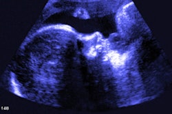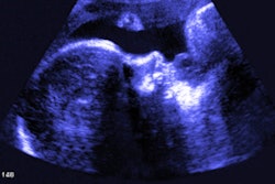
Ultrasound can be used to measure fetal growth alterations in diabetic women who struggle with controlling their blood glucose levels, according to Chinese research published April 7 in JAMA Network Open.
A team led by Jiao-jiao Zou from Fudan University in Shanghai found that pregnant women who received a diagnosis of gestational diabetes in mid-pregnancy or had hyperglycemia during all three trimesters showed an association with fetal overgrowth.
"It may be necessary to rethink when to screen for gestational diabetes in order to take precautions in early pregnancy to prevent excessive fetal growth," Zou and colleagues wrote.
Glucose is a major part of the maternal intrauterine environment. Previous research suggests that an intrauterine environment with high glucose levels can lead to long-term effects by altering an offspring's genetics and morphological characteristics and function.
Hyperglycemia and gestational diabetes create such conditions in the intrauterine environment, with research showing the latter affecting more than 10% of pregnancy outcomes. The researchers wrote that metabolic levels of blood glucose may be linked to growth and development during specific periods of pregnancy.
Zou and colleagues wanted to find the link between maternal blood glucose levels and fetal intrauterine growth at different gestational periods. Sonographers measured fetal biometrics using multiple ultrasonographic values (Voluson E8, GE Healthcare).
They looked at data from 3,746 women who had glycemic trajectory data and an average age of 28.6 years. Out of these, 983 women had gestational diabetes.
The women were also split into three trajectory groups: consistently normal glucose levels (3,566 women), hyperglycemia only in late pregnancy (101 women), and hyperglycemia in all three trimesters (79 women).
The study authors found that except for biparietal diameter, fetal biometrics as measured with ultrasound for the women with gestational diabetes or hyperglycemia in all three trimesters were significantly higher. This includes an estimated increase in fetal weight in the group with gestational diabetes (β = 1.82) and in women with hyperglycemia for all three trimesters (β = 1.50; p = 0.002).
Ultrasound also showed fetal biometric alterations among women with gestational diabetes appeared before 24 weeks' gestational age. The birth weight of newborns was on average 40.4 grams heavier than in women without gestational diabetes at 40.4 grams. The risk of large size for gestational age and macrosomia also increased with odds ratios of 1.36 and 1.47, respectively.
However, pregnant women with hyperglycemia only in late pregnancy showed significantly reduced fetal biometrics, with abdominal circumference increasing after 34 weeks' gestational age (β = 1.92).
The authors wrote that their results suggest that pregnant women, particularly those with high blood glucose levels throughout pregnancy should make efforts to prevent and control gestational diabetes, as well as avoid elevated blood glucose levels in late pregnancy.
"Monitoring the blood glucose level throughout pregnancy was a crucial component in assessing offspring growth patterns, especially for those pregnant women who did not receive a diagnosis of gestational diabetes but who had hyperglycemia in late pregnancy, because their offspring had significantly increased abdominal circumference after 34 weeks' gestational age," they added.