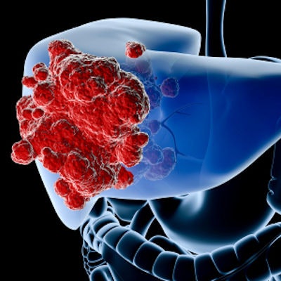
Ultrasound screenings with shorter intervals for high-risk patients with hepatocellular carcinoma (HCC) may save more lives and improve quality of life, according to research published June 24 in JAMA Network Open.
The results suggest a relatively easy way to improve HCC patient outcomes, wrote a team led by Dr. Shih-Chiang Kuo from the National Cheng Kung University Hospital in Taiwan.
"Our finding shows a clear indication of saving life-years and quality of life, and clinicians can use this knowledge to remind their patients, especially those with viral infection and/or cirrhosis of liver," said Dr. Jung-Der Wang, one of the study's co-authors.
Hepatocellular carcinoma accounts for most liver cancer cases. But despite improvements in medical technology and treatment, the five-year survival rate of HCC remains low, according to Kuo's team.
Early detection by screening is recommended by medical societies, but some research does not support six-month screening intervals using abdominal ultrasonography. Along with that, an "inadequate" number of high-risk patients do not adhere to regular screenings, the authors noted.
"Many patients with viral hepatitis B or C infection, or, cirrhosis of liver might feel tired of such examinations after negative results of the initial several years," Wang told AuntMinnie.com. "Then, many of them would consider that they might not be the unlucky one who would later develop HCC and stop the biannual screening ultrasonography."
Kuo and colleagues sought to investigate the optimal interval of ultrasonography screening for early diagnosis of HCC among high-risk patients via a study that included 59,194 patients; of these, 42,081 were men (average age, 62.2 years) and 17,113 were women (average age, 69).
The team found the longer the interval between ultrasonography screenings, the higher the loss of life expectancy and loss of quality-adjusted life expectancy for both men and women.
| Loss of quality-adjusted life years by ultrasound screening interval period in patients with HCC | |||||
| Gender | 6 months | 12 months | 24 months | 36 months | More than 36 months |
| Men | 10 | 11.1 | 12.1 | 13.1 | 14.6 |
| Women | 9 | 9.7 | 10.3 | 10.7 | 11.4 |
The authors also noted that patients with underlying hepatitis B virus infection or cirrhosis had the greatest improvement in life expectancy with shorter screening intervals.
Although multiple medical societies recommend ultrasonography screening for HCC every six to 12 months, the researchers found that only 31.4% of men and 42.2% of women with HCC in Taiwan underwent ultrasonography within six months before diagnosis, and only 39.3% of men and 51.9% of women underwent such screening within 12 months.
"Moreover, among those with underlying liver cirrhosis, only 35.5% of men and 49.1% of women underwent ultrasonography within six months before HCC diagnosis, indicating underutilization of ultrasonography screening in Taiwan," the team wrote.
The study highlights the fact that use of ultrasound for early detection of HCC could be improved, according to the authors.
"The results of this study suggest that regular ultrasonography screening with an interval of six to 12 months or less may lead to early detection of HCC and may save lives and improve utility for patients with HCC from a lifetime perspective," they concluded. "Because people with underlying risk factors ... showed only slightly more frequent ultrasonography screening than those without ... we recommend improving this clinical practice."
Wang told AuntMinnie.com that the team is looking into what even more frequent screening, namely three months, would do for patients, as well as the cost-effectiveness of such screening in terms of cost per quality-adjusted life-year saved.

![Examples of ultrasound findings and techniques. (A) Images in a 39-year-old male patient with a mass in the left thigh. The mass is heterogeneous on the B-mode US image (compared with the patient in D) and showed increased microvascularity (superb microvascular imaging [SMI]) and shear-wave elastography (SWE) values. Undifferentiated pleomorphic sarcoma was diagnosed at biopsy (with pleomorphic rhabdomyosarcoma in surgical specimen). (B) Images in an 18-year-old male patient with a mass in the left leg. The mass is hypoechoic on the B-mode image, with no other findings suggestive of malignancy. The lesion is in contact with the cortex of the tibia, which is slightly irregular. CT revealed a doubtful anteromedial tibial erosion. The microvascular study demonstrated high vascularization, suggestive of malignancy. Periosteal Ewing sarcoma was diagnosed with both histologic and immunohistochemical confirmation. (C) Images in a 69-year-old female patient with a lump growing on the outside of the left leg. Multiple SWE examinations were performed (please note the high values obtained in the measurements, whereas the color map highlights the stiffness relative to adjacent tissues). SMI showed areas of increased vascularization to target for sampling. Undifferentiated spindle cell sarcoma was diagnosed at biopsy, with residual leiomyosarcoma in the surgical specimen after neoadjuvant therapy. (D) Images in a 56-year-old female patient with a mass in the right thigh. The mass is heterogeneous at both B-mode ultrasound (similar to patient A) and MRI (coronal T2-weighted spectral attenuated inversion recovery [SPAIR]; T1-weighted pre-contrast and postcontrast imaging), which even shows uptake after the administration of paramagnetic contrast material, which is traditionally suggestive of malignancy. Low values at SMI and elastography are suggestive of benignity. Spindle cell lipoma was diagnosed at biopsy, with atypical spindle cell lipomatous tumor in the surgical specimen.](https://img.auntminnie.com/mindful/smg/workspaces/default/uploads/2026/08/images-radiol250278fig2.APCFLSvX6p.jpg?auto=format%2Ccompress&fit=crop&h=100&q=70&w=100)


![Examples of ultrasound findings and techniques. (A) Images in a 39-year-old male patient with a mass in the left thigh. The mass is heterogeneous on the B-mode US image (compared with the patient in D) and showed increased microvascularity (superb microvascular imaging [SMI]) and shear-wave elastography (SWE) values. Undifferentiated pleomorphic sarcoma was diagnosed at biopsy (with pleomorphic rhabdomyosarcoma in surgical specimen). (B) Images in an 18-year-old male patient with a mass in the left leg. The mass is hypoechoic on the B-mode image, with no other findings suggestive of malignancy. The lesion is in contact with the cortex of the tibia, which is slightly irregular. CT revealed a doubtful anteromedial tibial erosion. The microvascular study demonstrated high vascularization, suggestive of malignancy. Periosteal Ewing sarcoma was diagnosed with both histologic and immunohistochemical confirmation. (C) Images in a 69-year-old female patient with a lump growing on the outside of the left leg. Multiple SWE examinations were performed (please note the high values obtained in the measurements, whereas the color map highlights the stiffness relative to adjacent tissues). SMI showed areas of increased vascularization to target for sampling. Undifferentiated spindle cell sarcoma was diagnosed at biopsy, with residual leiomyosarcoma in the surgical specimen after neoadjuvant therapy. (D) Images in a 56-year-old female patient with a mass in the right thigh. The mass is heterogeneous at both B-mode ultrasound (similar to patient A) and MRI (coronal T2-weighted spectral attenuated inversion recovery [SPAIR]; T1-weighted pre-contrast and postcontrast imaging), which even shows uptake after the administration of paramagnetic contrast material, which is traditionally suggestive of malignancy. Low values at SMI and elastography are suggestive of benignity. Spindle cell lipoma was diagnosed at biopsy, with atypical spindle cell lipomatous tumor in the surgical specimen.](https://img.auntminnie.com/mindful/smg/workspaces/default/uploads/2026/08/images-radiol250278fig2.APCFLSvX6p.jpg?auto=format%2Ccompress&fit=crop&h=112&q=70&w=112)