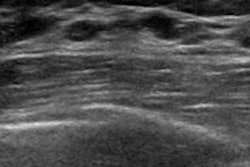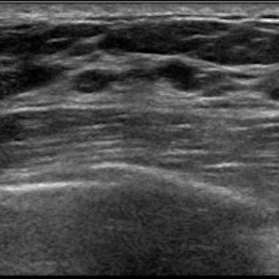
Artificial intelligence (AI) can detect and diagnose cancer on breast ultrasound with accuracy comparable to that of radiologists, according to research presented Monday at the 2021 virtual annual meeting of the American Roentgen Ray Society (ARRS).
A research team from New York University trained its own AI algorithm to automatically detect and classify breast lesions on ultrasound without manual annotation from radiologists. The team also said that AI systems such as this can help human imagers make more accurate diagnoses.
"We believe this work highlights the promise that deep learning and AI might hold for improving breast cancer detection using breast ultrasound," said Jamie Oliver, a medical student at New York University.
Breast ultrasound is an important tool in the detection of breast masses. While studies have shown that the technique can consistently detect additional cancers when used as a supplemental screening modality, it is prone to false positives and has lower sensitivity compared with breast MRI.
The NYU team trained its AI model using its large-scale, institutional dataset of 345,370 breast ultrasound exams acquired from 168,282 patients between 2012 and 2019.
The team then tested the algorithm alongside 13 human imaging readers with varied expertise. Each reader reviewed 250 breast ultrasound exams that were randomly sampled from the test set. A hybrid decision-making model was then created for each reader, which made predictions by evenly weighting the predictions of the reader and the AI system.
Among the exams included in the study, 392 breasts were imaged, 38 of which had malignant lesions.
The AI model achieved an area under the receiver operating characteristic (AUC) of 0.880, significantly outperforming each of the 13 readers (p < 0.01). The performances of the human readers achieved 0.818 AUC on average, none of which exceeded the AI model's performance.
However, the accuracy of the readers improved with the help of AI. The hybrid predictions of each reader and the AI algorithm improved the average diagnostic accuracy for all readers, including attending radiologists and trainees, to 0.852 AUC (p < 0.01).
| Accuracy of breast ultrasound with and without AI assistance | ||
| Average AUC without AI assistance | Average AUC with AI assistance | |
| Radiologists | 0.824 | 0.863 |
| Trainees | 0.805 | 0.826 |
"If incorporated as a decision-support tool, we believe that AI has the potential to improve the diagnostic accuracy of breast ultrasound and significantly decrease the number of benign biopsies performed," Oliver said.
The team is also optimistic that such an AI system can serve as a valuable aid in clinical decision-making in areas with reduced resources, including where there might be shortages of radiologists.


![Examples of ultrasound findings and techniques. (A) Images in a 39-year-old male patient with a mass in the left thigh. The mass is heterogeneous on the B-mode US image (compared with the patient in D) and showed increased microvascularity (superb microvascular imaging [SMI]) and shear-wave elastography (SWE) values. Undifferentiated pleomorphic sarcoma was diagnosed at biopsy (with pleomorphic rhabdomyosarcoma in surgical specimen). (B) Images in an 18-year-old male patient with a mass in the left leg. The mass is hypoechoic on the B-mode image, with no other findings suggestive of malignancy. The lesion is in contact with the cortex of the tibia, which is slightly irregular. CT revealed a doubtful anteromedial tibial erosion. The microvascular study demonstrated high vascularization, suggestive of malignancy. Periosteal Ewing sarcoma was diagnosed with both histologic and immunohistochemical confirmation. (C) Images in a 69-year-old female patient with a lump growing on the outside of the left leg. Multiple SWE examinations were performed (please note the high values obtained in the measurements, whereas the color map highlights the stiffness relative to adjacent tissues). SMI showed areas of increased vascularization to target for sampling. Undifferentiated spindle cell sarcoma was diagnosed at biopsy, with residual leiomyosarcoma in the surgical specimen after neoadjuvant therapy. (D) Images in a 56-year-old female patient with a mass in the right thigh. The mass is heterogeneous at both B-mode ultrasound (similar to patient A) and MRI (coronal T2-weighted spectral attenuated inversion recovery [SPAIR]; T1-weighted pre-contrast and postcontrast imaging), which even shows uptake after the administration of paramagnetic contrast material, which is traditionally suggestive of malignancy. Low values at SMI and elastography are suggestive of benignity. Spindle cell lipoma was diagnosed at biopsy, with atypical spindle cell lipomatous tumor in the surgical specimen.](https://img.auntminnie.com/mindful/smg/workspaces/default/uploads/2026/08/images-radiol250278fig2.APCFLSvX6p.jpg?auto=format%2Ccompress&fit=crop&h=100&q=70&w=100)


![Examples of ultrasound findings and techniques. (A) Images in a 39-year-old male patient with a mass in the left thigh. The mass is heterogeneous on the B-mode US image (compared with the patient in D) and showed increased microvascularity (superb microvascular imaging [SMI]) and shear-wave elastography (SWE) values. Undifferentiated pleomorphic sarcoma was diagnosed at biopsy (with pleomorphic rhabdomyosarcoma in surgical specimen). (B) Images in an 18-year-old male patient with a mass in the left leg. The mass is hypoechoic on the B-mode image, with no other findings suggestive of malignancy. The lesion is in contact with the cortex of the tibia, which is slightly irregular. CT revealed a doubtful anteromedial tibial erosion. The microvascular study demonstrated high vascularization, suggestive of malignancy. Periosteal Ewing sarcoma was diagnosed with both histologic and immunohistochemical confirmation. (C) Images in a 69-year-old female patient with a lump growing on the outside of the left leg. Multiple SWE examinations were performed (please note the high values obtained in the measurements, whereas the color map highlights the stiffness relative to adjacent tissues). SMI showed areas of increased vascularization to target for sampling. Undifferentiated spindle cell sarcoma was diagnosed at biopsy, with residual leiomyosarcoma in the surgical specimen after neoadjuvant therapy. (D) Images in a 56-year-old female patient with a mass in the right thigh. The mass is heterogeneous at both B-mode ultrasound (similar to patient A) and MRI (coronal T2-weighted spectral attenuated inversion recovery [SPAIR]; T1-weighted pre-contrast and postcontrast imaging), which even shows uptake after the administration of paramagnetic contrast material, which is traditionally suggestive of malignancy. Low values at SMI and elastography are suggestive of benignity. Spindle cell lipoma was diagnosed at biopsy, with atypical spindle cell lipomatous tumor in the surgical specimen.](https://img.auntminnie.com/mindful/smg/workspaces/default/uploads/2026/08/images-radiol250278fig2.APCFLSvX6p.jpg?auto=format%2Ccompress&fit=crop&h=112&q=70&w=112)