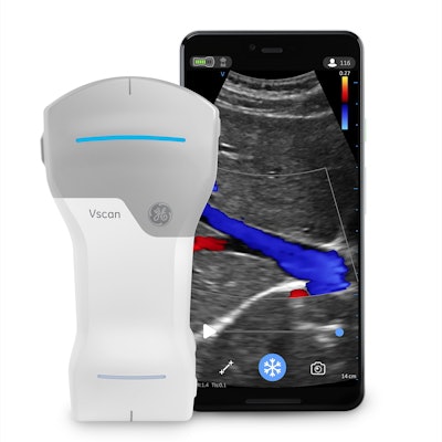
GE Healthcare has unveiled Vscan Air, a new compact ultrasound system designed to aid in the assessment of COVID-19 patients and to enable primary care physicians to perform whole-body ultrasound scanning. It's available now in the U.S. and Europe.
A pocket-sized wireless system that's powered by a mobile app, Vscan Air features two piezoelectric transducers in a single-probe design. Clinicians can utilize a high-frequency linear transducer for shallow scanning or a curved transducer for deep scanning -- just by flipping the probe around, according to the vendor.
GE is also highlighting the image quality of Vscan Air; the company said it utilized its SignalMax technology to adapt signal-processing methods from its higher-end ultrasound systems.
GE's new Vscan Air compact ultrasound unit. All images and video clips courtesy of GE.The launch of Vscan Air comes at a time when ultrasound is increasingly being utilized at the bedside. The COVID-19 pandemic has resulted in clinicians treating more critically ill patients than ever before, and with limited staff, time, and resources, said Anders Wold, CEO of ultrasound at GE.
"And they're now looking for smaller and more mobile technology at the point of care to address the growing list of critical medical needs, quickly and efficiently," he said.
In addition, clinicians are changing how they are providing care, Wold said. Healthcare is decentralizing and moving outside of hospitals.
"Therefore, this shift toward smaller, portable, and easy-to-use devices that are more easily deployed across a variety of uses has been underway for several years," he said. "COVID-19 has accelerated these trends, and the demand for ultrasound -- particularly handheld ultrasound -- is surging. We have seen massive demand for these devices over the last year in particular."
Clinicians have used the device to scan the heart and lungs, triaging patients quickly and confidently, he said.
Abnormal lung image with B-lines acquired with Vscan Air.
Healthy parasternal color cardiac image produced by Vscan Air.
"Fighting COVID-19 is important, but our purpose is to transform the physical exam by putting personalized care and immediate visualization in the pockets of clinicians worldwide," he said.
With the latest generation of Vscan handheld ultrasound systems, GE wanted to broaden the usage of its technology beyond the highly specialized branches of medicine such as obstetrics or cardiology and support primary care physicians, said Steffen Mueller, Vscan product manager.
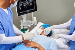
The wireless probe also confers an ergonomic advantage during needle procedures, as clinicians don't have to worry about wires breaching the sterile environment, according to the vendor. The probe is waterproof with an IP 67 rating and can be fully immersed in cleaning agents. It's also protected from dust, moisture, and grime, GE said.
Images or videos can be shared with clinicians or colleagues in a variety of methods, including AirDrop, texting, or emailing. They're also automatically anonymized for patient data security, GE said.



![Examples of ultrasound findings and techniques. (A) Images in a 39-year-old male patient with a mass in the left thigh. The mass is heterogeneous on the B-mode US image (compared with the patient in D) and showed increased microvascularity (superb microvascular imaging [SMI]) and shear-wave elastography (SWE) values. Undifferentiated pleomorphic sarcoma was diagnosed at biopsy (with pleomorphic rhabdomyosarcoma in surgical specimen). (B) Images in an 18-year-old male patient with a mass in the left leg. The mass is hypoechoic on the B-mode image, with no other findings suggestive of malignancy. The lesion is in contact with the cortex of the tibia, which is slightly irregular. CT revealed a doubtful anteromedial tibial erosion. The microvascular study demonstrated high vascularization, suggestive of malignancy. Periosteal Ewing sarcoma was diagnosed with both histologic and immunohistochemical confirmation. (C) Images in a 69-year-old female patient with a lump growing on the outside of the left leg. Multiple SWE examinations were performed (please note the high values obtained in the measurements, whereas the color map highlights the stiffness relative to adjacent tissues). SMI showed areas of increased vascularization to target for sampling. Undifferentiated spindle cell sarcoma was diagnosed at biopsy, with residual leiomyosarcoma in the surgical specimen after neoadjuvant therapy. (D) Images in a 56-year-old female patient with a mass in the right thigh. The mass is heterogeneous at both B-mode ultrasound (similar to patient A) and MRI (coronal T2-weighted spectral attenuated inversion recovery [SPAIR]; T1-weighted pre-contrast and postcontrast imaging), which even shows uptake after the administration of paramagnetic contrast material, which is traditionally suggestive of malignancy. Low values at SMI and elastography are suggestive of benignity. Spindle cell lipoma was diagnosed at biopsy, with atypical spindle cell lipomatous tumor in the surgical specimen.](https://img.auntminnie.com/mindful/smg/workspaces/default/uploads/2026/08/images-radiol250278fig2.APCFLSvX6p.jpg?auto=format%2Ccompress&fit=crop&h=100&q=70&w=100)


![Examples of ultrasound findings and techniques. (A) Images in a 39-year-old male patient with a mass in the left thigh. The mass is heterogeneous on the B-mode US image (compared with the patient in D) and showed increased microvascularity (superb microvascular imaging [SMI]) and shear-wave elastography (SWE) values. Undifferentiated pleomorphic sarcoma was diagnosed at biopsy (with pleomorphic rhabdomyosarcoma in surgical specimen). (B) Images in an 18-year-old male patient with a mass in the left leg. The mass is hypoechoic on the B-mode image, with no other findings suggestive of malignancy. The lesion is in contact with the cortex of the tibia, which is slightly irregular. CT revealed a doubtful anteromedial tibial erosion. The microvascular study demonstrated high vascularization, suggestive of malignancy. Periosteal Ewing sarcoma was diagnosed with both histologic and immunohistochemical confirmation. (C) Images in a 69-year-old female patient with a lump growing on the outside of the left leg. Multiple SWE examinations were performed (please note the high values obtained in the measurements, whereas the color map highlights the stiffness relative to adjacent tissues). SMI showed areas of increased vascularization to target for sampling. Undifferentiated spindle cell sarcoma was diagnosed at biopsy, with residual leiomyosarcoma in the surgical specimen after neoadjuvant therapy. (D) Images in a 56-year-old female patient with a mass in the right thigh. The mass is heterogeneous at both B-mode ultrasound (similar to patient A) and MRI (coronal T2-weighted spectral attenuated inversion recovery [SPAIR]; T1-weighted pre-contrast and postcontrast imaging), which even shows uptake after the administration of paramagnetic contrast material, which is traditionally suggestive of malignancy. Low values at SMI and elastography are suggestive of benignity. Spindle cell lipoma was diagnosed at biopsy, with atypical spindle cell lipomatous tumor in the surgical specimen.](https://img.auntminnie.com/mindful/smg/workspaces/default/uploads/2026/08/images-radiol250278fig2.APCFLSvX6p.jpg?auto=format%2Ccompress&fit=crop&h=112&q=70&w=112)