A two-and-a-half-hour online course boosted the knowledge of COVID-19 lung ultrasound presentation among ob/gyn clinicians with no prior lung ultrasound experience, according to the findings of a small study published on April 29 in Ultrasound in Obstetrics & Gynaecology.
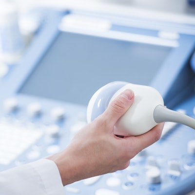
While many ob/gyn physicians have ultrasound equipment and experience for performing prenatal exams, they're to have little to no experience using those tools to perform lung ultrasound scans. However, the new research demonstrates that ob/gyns may be able to quickly pick up the skills needed to distinguish normal findings from those that might suggest a patient has the novel coronavirus.
"We believe that our course represents one possible model of implementing lung ultrasound education, and it might be a useful launchpad for gynecologists/obstetricians involved in the management of COVID-19 pregnant patients," wrote the team of Italian authors, led by Danilo Buonsenso, PhD, and Dr. Francesca Moro, PhD, from Fondazione Policlinico Universitario Agostino Gemelli in Rome.
Lung ultrasound scans have proved to be an effective, noninvasive modality for screening patients, including pregnant patients, with confirmed or suspected cases of the novel coronavirus. The latest study adds to existing research demonstrating ob/gyn clinicians may be well-poised to recognize B-lines, subpleural consolidations, a thickened or patchy pleural line, and other hallmark signs of potential infection with the novel coronavirus.
For their study, Buonsenso and Moro recruited two pneumologists and one pediatrician, each with more than 10 years of lung ultrasound experience, to create an online training module.
The module begins with a 15-minute pretest that includes 10 ultrasound video clips showing healthy, normal lungs or ones with pathological patterns. The test asks participants to choose whether the lung video shows the following:
a) Normal findings
b) Clear and distinct vertical artifacts with small pleural alterations
c) Broken pleural line and small consolidations
d) White lung without evidence subpleural consolidations
e) White lung with evidence subpleural consolidations
f) Pattern not compatible, vast consolidations
g) Pattern not compatible, pleural effusions
Following the pretest, participants watch a two-hour theoretical course providing lung ultrasound education. Finally, the course ends with a 15-minute test showing the same 10 ultrasound clips.
The researchers tested the training module's effectiveness with 11 ob/gyn clinicians who had no prior lung ultrasound experience. The clinicians' median number of correct answers rose from 6/10 at the pretest to 9/10 at the final test.
In addition, eight clinicians correctly distinguished between normal and pathological lung findings in all presented cases. Another two clinicians missed one answer, and one missed two answers.
Although the study was small, the authors believe the module could help quickly train ob/gyn clinicians who may need to see and manage pregnant patients with COVID-19.
"In conclusion, our preliminary experience show that a fast teaching program was sufficient to provide appropriate theoretical skills to recognize specific lung ultrasound patterns," they wrote.