When a pregnant woman sees her fetus on imaging scans, it can help strengthen the bond she feels with her unborn child, say researchers. They demonstrate how 3D-printed models based on ultrasound scans confirmed this concept in a study published online on February 8 in the Journal of Obstetric, Gynecologic and Neonatal Nursing.
Maternal-fetal attachment describes the emotional bond that typically develops between a pregnant woman and her unborn child. The strength of this bond is likely associated with the health or behavior of the mother and the well-being of the child, with higher levels of attachment generally resulting in greater degrees of postnatal bonding, noted Dr. John Coté and colleagues from Creighton University School of Medicine.
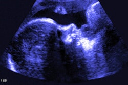
Ultrasound exams are routinely performed during maternity care and showing these scans to mothers has been associated with increased maternal-fetal attachment, the authors continued. Preliminary studies have also suggested that the use of more advanced modalities could further increase attachment levels.
To explore this possibility, Coté and colleagues conducted a randomized controlled trial in which they assessed maternal-fetal attachment levels in 96 pregnant women. All the women underwent an ultrasound exam during the third trimester of pregnancy.
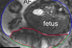
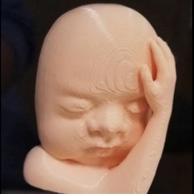
To create the 3D-printed models, the researchers extracted and processed ultrasound scans of the fetuses, converted the files into virtual 3D models, and then sent the 3D models to a 3D printer. The resulting fetal figurines served as representations of the fetus based on the ultrasound data and were not exact replicas.
 3D-printed model of a fetus' face based on ultrasound scans. Image courtesy of Amy Brack, PhD.
To assess bonding, each of the study participants completed a Maternal Antenatal Attachment Scale (MAAS) questionnaire, the primary tool used to assess maternal-fetal attachment. Questionnaires were completed once before undergoing the ultrasound exam and a second time two weeks after receiving either an ultrasound scan alone or an ultrasound scan plus a 3D-printed model of the fetus. MAAS measures the quality of 19 different aspects of maternal-fetal attachment on a five-point Likert-type scale.
For both groups of mothers, the increases in average MAAS scores before and after receiving the ultrasound scan and/or 3D-printed model were statistically significant, from 76.5 to 83.4 points (out of a max score of 95).
In comparing the two groups, however, the researchers found that the increase in average MAAS score was nearly twofold greater for the 3D printing cohort than the ultrasound-only cohort. The average MAAS score increased by 8.7 points for the 3D printing cohort, compared with only 4.9 points for the ultrasound-only cohort -- a statistically significant difference (p = 0.002).
"We propose the use of advanced ultrasonography and 3D prints, not to medicalize pregnancy, but rather, to address whole-person needs by providing women tangible ways to visualize their fetuses during pregnancy, enhance prenatal bonding, and provide women with new ways to share their pregnancy experiences with their social support networks," the authors wrote.