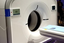
The parent company of Philips Healthcare has filed a lawsuit against an imaging equipment service firm in the state of Washington, charging the company with hacking proprietary Philips software for ultrasound scanners in order to activate features and enable upgrades that users haven't paid for.
The litigation charges Summit Imaging and its CEO, Larry Nguyen, with a "fraudulent and intentional scheme" to circumvent access controls Philips has placed on its ultrasound scanners, arguing that Summit hacked the access controls with the goal of supporting its repair parts and service business. The lawsuit was filed on October 29 in the U.S. District Court for the Western District of Washington.
The complaint filed in the case notes that all of Philips' ultrasound scanners run on one of two software platforms, one called Philips Voyager Platform and the other called Philips Common Platform. The company's software includes access controls that limit access to some features and also control access to optional applications that enable additional system features for which Philips charges end users.
Further, Philips uses "multiple layers of technological controls" to protect its software from unauthorized access. These controls include user-specific access codes and hardware keys that are customized for particular end users. These controls extend to ultrasound transducers, which will only function on a particular scanner if the software has been enabled.
The complaint charges that Summit Imaging sells customers service and repair contracts for ultrasound scanners and markets its repair and service capabilities "in ways that fall well outside the bounds of standard clinical user level access."
Specifically, the complaint charges that Summit created a software program called Adepto that was "specifically designed to hack into Philips medical imaging devices by circumventing Philips' access controls. Calling Adepto a "hacking tool," the complaint charges that Summit markets the software as a "legal solution" for customers that are "aware of the issues surrounding licensing copyright" and as "the only legal alternative to OEM service."
The complaint alleges that on service calls, Summit personnel physically remove the hard drive from a Philips ultrasound scanner and attach it to a separate computer that's running the Adepto software. Adepto then bypasses access controls set by Philips software and accesses the removed hard drive. Adepto is then able to install copies of Philips software with unlicensed options on it, the complaint alleges.
What's more, Summit Imaging provides customers with copies of Adepto and training on how to use it, enabling them to "repeatedly hack into and circumvent Philips' access controls in order to gain access to unlicensed access to Philips' copyright-protected material."
The complaint ends by charging Nguyen and Summit Imaging with a variety of charges, including trade secret misappropriation, unfair competition, modifying copyright management information, circumventing a technological measure, and false advertising. The complaint asks that Summit be prohibited from engaging in activities that are covered in the complaint and that Philips be awarded damages for lost profits, as well as compensatory and punitive damages.

![Examples of ultrasound findings and techniques. (A) Images in a 39-year-old male patient with a mass in the left thigh. The mass is heterogeneous on the B-mode US image (compared with the patient in D) and showed increased microvascularity (superb microvascular imaging [SMI]) and shear-wave elastography (SWE) values. Undifferentiated pleomorphic sarcoma was diagnosed at biopsy (with pleomorphic rhabdomyosarcoma in surgical specimen). (B) Images in an 18-year-old male patient with a mass in the left leg. The mass is hypoechoic on the B-mode image, with no other findings suggestive of malignancy. The lesion is in contact with the cortex of the tibia, which is slightly irregular. CT revealed a doubtful anteromedial tibial erosion. The microvascular study demonstrated high vascularization, suggestive of malignancy. Periosteal Ewing sarcoma was diagnosed with both histologic and immunohistochemical confirmation. (C) Images in a 69-year-old female patient with a lump growing on the outside of the left leg. Multiple SWE examinations were performed (please note the high values obtained in the measurements, whereas the color map highlights the stiffness relative to adjacent tissues). SMI showed areas of increased vascularization to target for sampling. Undifferentiated spindle cell sarcoma was diagnosed at biopsy, with residual leiomyosarcoma in the surgical specimen after neoadjuvant therapy. (D) Images in a 56-year-old female patient with a mass in the right thigh. The mass is heterogeneous at both B-mode ultrasound (similar to patient A) and MRI (coronal T2-weighted spectral attenuated inversion recovery [SPAIR]; T1-weighted pre-contrast and postcontrast imaging), which even shows uptake after the administration of paramagnetic contrast material, which is traditionally suggestive of malignancy. Low values at SMI and elastography are suggestive of benignity. Spindle cell lipoma was diagnosed at biopsy, with atypical spindle cell lipomatous tumor in the surgical specimen.](https://img.auntminnie.com/mindful/smg/workspaces/default/uploads/2026/08/images-radiol250278fig2.APCFLSvX6p.jpg?auto=format%2Ccompress&fit=crop&h=100&q=70&w=100)


![Examples of ultrasound findings and techniques. (A) Images in a 39-year-old male patient with a mass in the left thigh. The mass is heterogeneous on the B-mode US image (compared with the patient in D) and showed increased microvascularity (superb microvascular imaging [SMI]) and shear-wave elastography (SWE) values. Undifferentiated pleomorphic sarcoma was diagnosed at biopsy (with pleomorphic rhabdomyosarcoma in surgical specimen). (B) Images in an 18-year-old male patient with a mass in the left leg. The mass is hypoechoic on the B-mode image, with no other findings suggestive of malignancy. The lesion is in contact with the cortex of the tibia, which is slightly irregular. CT revealed a doubtful anteromedial tibial erosion. The microvascular study demonstrated high vascularization, suggestive of malignancy. Periosteal Ewing sarcoma was diagnosed with both histologic and immunohistochemical confirmation. (C) Images in a 69-year-old female patient with a lump growing on the outside of the left leg. Multiple SWE examinations were performed (please note the high values obtained in the measurements, whereas the color map highlights the stiffness relative to adjacent tissues). SMI showed areas of increased vascularization to target for sampling. Undifferentiated spindle cell sarcoma was diagnosed at biopsy, with residual leiomyosarcoma in the surgical specimen after neoadjuvant therapy. (D) Images in a 56-year-old female patient with a mass in the right thigh. The mass is heterogeneous at both B-mode ultrasound (similar to patient A) and MRI (coronal T2-weighted spectral attenuated inversion recovery [SPAIR]; T1-weighted pre-contrast and postcontrast imaging), which even shows uptake after the administration of paramagnetic contrast material, which is traditionally suggestive of malignancy. Low values at SMI and elastography are suggestive of benignity. Spindle cell lipoma was diagnosed at biopsy, with atypical spindle cell lipomatous tumor in the surgical specimen.](https://img.auntminnie.com/mindful/smg/workspaces/default/uploads/2026/08/images-radiol250278fig2.APCFLSvX6p.jpg?auto=format%2Ccompress&fit=crop&h=112&q=70&w=112)