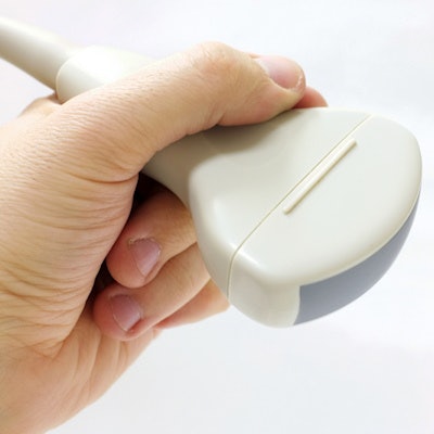
A Pennsylvania hospital is facing legal action due to the discovery that improperly cleaned ultrasound probes were used on more than 200 patients, according to a report from CBS affiliate KDKA 2 in Pittsburgh.
Pittsburgh attorney Dallas Hartman is bringing the suit against UPMC Jameson of New Castle on behalf of patients who were exposed to dirty probes as far back as October 2017, KDKA 2 said.
The action comes after an internal review conducted by the hospital found that ultrasound probes were improperly cleaned, according to another article about the situation posted on November 2 by the Pittsburgh Post-Gazette. Hundreds of patients who underwent ultrasound scans for prostate or ob/gyn exams may have been affected, the Post-Gazette said.
UPMC Jameson acknowledged that proper cleaning processes were not documented and followed for some probes used for internal prostate and ob/gyn exams, but added that any health risks were low and corrective measure were implemented, KDKA 2 noted. Also, the hospital alerted both the state Department of Health and the Joint Commission.
"In an abundance of caution, we have contacted all potentially affected patients with additional information, offering free precautionary blood and urine testing to ensure patient safety," the hospital noted. "UPMC Jameson is committed to providing high-quality care in the safest environment possible. We apologize for the concern and inconvenience this matter has caused our patients and their families."








![Examples of ultrasound findings and techniques. (A) Images in a 39-year-old male patient with a mass in the left thigh. The mass is heterogeneous on the B-mode US image (compared with the patient in D) and showed increased microvascularity (superb microvascular imaging [SMI]) and shear-wave elastography (SWE) values. Undifferentiated pleomorphic sarcoma was diagnosed at biopsy (with pleomorphic rhabdomyosarcoma in surgical specimen). (B) Images in an 18-year-old male patient with a mass in the left leg. The mass is hypoechoic on the B-mode image, with no other findings suggestive of malignancy. The lesion is in contact with the cortex of the tibia, which is slightly irregular. CT revealed a doubtful anteromedial tibial erosion. The microvascular study demonstrated high vascularization, suggestive of malignancy. Periosteal Ewing sarcoma was diagnosed with both histologic and immunohistochemical confirmation. (C) Images in a 69-year-old female patient with a lump growing on the outside of the left leg. Multiple SWE examinations were performed (please note the high values obtained in the measurements, whereas the color map highlights the stiffness relative to adjacent tissues). SMI showed areas of increased vascularization to target for sampling. Undifferentiated spindle cell sarcoma was diagnosed at biopsy, with residual leiomyosarcoma in the surgical specimen after neoadjuvant therapy. (D) Images in a 56-year-old female patient with a mass in the right thigh. The mass is heterogeneous at both B-mode ultrasound (similar to patient A) and MRI (coronal T2-weighted spectral attenuated inversion recovery [SPAIR]; T1-weighted pre-contrast and postcontrast imaging), which even shows uptake after the administration of paramagnetic contrast material, which is traditionally suggestive of malignancy. Low values at SMI and elastography are suggestive of benignity. Spindle cell lipoma was diagnosed at biopsy, with atypical spindle cell lipomatous tumor in the surgical specimen.](https://img.auntminnie.com/mindful/smg/workspaces/default/uploads/2026/08/images-radiol250278fig2.APCFLSvX6p.jpg?auto=format%2Ccompress&fit=crop&h=112&q=70&w=112)


