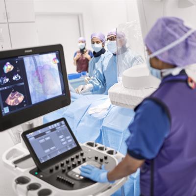
Royal Philips, the parent company of Philips Healthcare, said it will introduce Epiq CVx, a line of new cardiovascular ultrasound scanners, at the upcoming European Society of Cardiology (ESC) meeting in Munich.
Epiq CVx is designed to provide improved image quality for cardiovascular imaging with a cardiology-centric design that enables clinicians to perform faster exams. It offers photorealistic 3D organ modeling with the company's TrueVue mode, which may help improve the diagnostic capability of echocardiography in different clinical scenarios, according to the company. Other features include image slicing and rapid quantification of cardiac anatomy.
Philips' Epiq CVx, a line of new cardiovascular ultrasound scanners.Also, the OLED monitor offers a more dynamic, wider viewing angle for side-by-side image comparison, according to the company. Philips believes that combining TrueVue with the OLED monitor will enable clinicians to offer better care, such as for pediatric patients.
A feature called Dynamic Heart Model uses anatomical intelligence to automatically quantify left ventricle function to produce a multibeat analysis for adult patients. The mode has been shown to reduce the amount of time needed to produce a 3D ejection fraction, and also delivers a high level of robustness and reliability, the company said.
Philips is also launching Epiq CVxi, another scanner in the CVx line for live imaging of interventional echocardiography procedures in catheterization labs. Epiq CVxi integrates ultrasound and angiography into a single system -- streamlining communication between the interventional cardiologist and the echocardiographer for structural heart procedures, Philips said.
Both Epiq CVx and Epiq CVxi have received the CE Mark and 510(k) clearance from the U.S. Food and Drug Administration (FDA).









![Examples of ultrasound findings and techniques. (A) Images in a 39-year-old male patient with a mass in the left thigh. The mass is heterogeneous on the B-mode US image (compared with the patient in D) and showed increased microvascularity (superb microvascular imaging [SMI]) and shear-wave elastography (SWE) values. Undifferentiated pleomorphic sarcoma was diagnosed at biopsy (with pleomorphic rhabdomyosarcoma in surgical specimen). (B) Images in an 18-year-old male patient with a mass in the left leg. The mass is hypoechoic on the B-mode image, with no other findings suggestive of malignancy. The lesion is in contact with the cortex of the tibia, which is slightly irregular. CT revealed a doubtful anteromedial tibial erosion. The microvascular study demonstrated high vascularization, suggestive of malignancy. Periosteal Ewing sarcoma was diagnosed with both histologic and immunohistochemical confirmation. (C) Images in a 69-year-old female patient with a lump growing on the outside of the left leg. Multiple SWE examinations were performed (please note the high values obtained in the measurements, whereas the color map highlights the stiffness relative to adjacent tissues). SMI showed areas of increased vascularization to target for sampling. Undifferentiated spindle cell sarcoma was diagnosed at biopsy, with residual leiomyosarcoma in the surgical specimen after neoadjuvant therapy. (D) Images in a 56-year-old female patient with a mass in the right thigh. The mass is heterogeneous at both B-mode ultrasound (similar to patient A) and MRI (coronal T2-weighted spectral attenuated inversion recovery [SPAIR]; T1-weighted pre-contrast and postcontrast imaging), which even shows uptake after the administration of paramagnetic contrast material, which is traditionally suggestive of malignancy. Low values at SMI and elastography are suggestive of benignity. Spindle cell lipoma was diagnosed at biopsy, with atypical spindle cell lipomatous tumor in the surgical specimen.](https://img.auntminnie.com/mindful/smg/workspaces/default/uploads/2026/08/images-radiol250278fig2.APCFLSvX6p.jpg?auto=format%2Ccompress&fit=crop&h=112&q=70&w=112)
