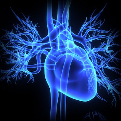
Echocardiography of pregnant women with pre-eclampsia has shown that the condition negatively affects the heart's ability to relax between contractions, leading to inefficient pumping and an overworked heart, according to a study published in the Journal of the American College of Cardiology.
The finding sheds light on why pre-eclampsia -- which boosts blood pressure and can cause organ damage -- increases the risk of heart failure, heart attack, and stroke in women who had it but recovered. The condition occurs in 5% to 8% of pregnant women, wrote a team led by Dr. Arthur Jason Vaught of Johns Hopkins University (JACC, July 3, 2018, Vol. 72:1).
Vaught and colleagues used echocardiography to evaluate the pumping and relaxing activity in the hearts of 63 women with severe pre-eclampsia as well as in 36 women without the condition. Severe pre-eclampsia was defined as blood pressure higher than 160/100 mm Hg, abnormal levels of protein in the urine, diagnosed kidney or liver damage, fluid in the lungs, low platelet counts, and vision problems.
All of the women underwent echocardiography at around 33 weeks of pregnancy. Those with severe pre-eclampsia had higher contraction pressures in the right ventricle of the heart; the researchers found a 5% difference in the change of the heart's shape in those with severe pre-eclampsia compared with healthy controls. Women with severe pre-eclampsia had abnormal periods between contractions, which decreased the heart's ability to pump blood and overworked the heart when it did, leading to heart failure, the group noted. Six of the women with severe pre-eclampsia also had peripartum pulmonary edema.
"The damage done to the heart's pumping ability during pregnancy in women with pre-eclampsia is striking, and it makes sense that this particular kind of damage puts them at greater risk of heart disease and strokes in the future," Vaught said in a statement released by Johns Hopkins.






![Examples of ultrasound findings and techniques. (A) Images in a 39-year-old male patient with a mass in the left thigh. The mass is heterogeneous on the B-mode US image (compared with the patient in D) and showed increased microvascularity (superb microvascular imaging [SMI]) and shear-wave elastography (SWE) values. Undifferentiated pleomorphic sarcoma was diagnosed at biopsy (with pleomorphic rhabdomyosarcoma in surgical specimen). (B) Images in an 18-year-old male patient with a mass in the left leg. The mass is hypoechoic on the B-mode image, with no other findings suggestive of malignancy. The lesion is in contact with the cortex of the tibia, which is slightly irregular. CT revealed a doubtful anteromedial tibial erosion. The microvascular study demonstrated high vascularization, suggestive of malignancy. Periosteal Ewing sarcoma was diagnosed with both histologic and immunohistochemical confirmation. (C) Images in a 69-year-old female patient with a lump growing on the outside of the left leg. Multiple SWE examinations were performed (please note the high values obtained in the measurements, whereas the color map highlights the stiffness relative to adjacent tissues). SMI showed areas of increased vascularization to target for sampling. Undifferentiated spindle cell sarcoma was diagnosed at biopsy, with residual leiomyosarcoma in the surgical specimen after neoadjuvant therapy. (D) Images in a 56-year-old female patient with a mass in the right thigh. The mass is heterogeneous at both B-mode ultrasound (similar to patient A) and MRI (coronal T2-weighted spectral attenuated inversion recovery [SPAIR]; T1-weighted pre-contrast and postcontrast imaging), which even shows uptake after the administration of paramagnetic contrast material, which is traditionally suggestive of malignancy. Low values at SMI and elastography are suggestive of benignity. Spindle cell lipoma was diagnosed at biopsy, with atypical spindle cell lipomatous tumor in the surgical specimen.](https://img.auntminnie.com/mindful/smg/workspaces/default/uploads/2026/08/images-radiol250278fig2.APCFLSvX6p.jpg?auto=format%2Ccompress&fit=crop&h=112&q=70&w=112)
