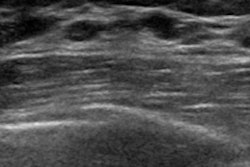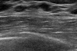
Computer-aided detection (CAD) software based on deep-learning technology can significantly reduce interpretation times for screening automated breast ultrasound (ABUS) studies without negatively affecting accuracy, according to research published in the August issue of the American Journal of Roentgenology.
In a retrospective observer performance study involving 18 radiologists and 185 screening ABUS studies, researchers led by Yulei Jiang, PhD, of the University of Chicago found that radiologists using CAD concurrently during ABUS interpretation could read the studies one-third faster than when they weren't using the software. Interpretation accuracy was comparable between the two methods.
"Use of the concurrent-read CAD system for interpretation of screening ABUS studies of women with dense breast tissue who do not have symptoms is expected to make interpretation significantly faster and produce noninferior diagnostic accuracy compared with interpretation without the CAD system," the authors wrote (AJR, August 2018, Vol. 211:2, pp. 452-461).
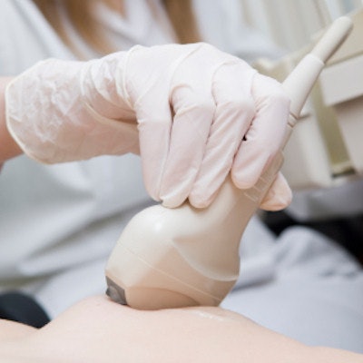
ABUS has been shown to be a useful adjunct to screening mammography for breast cancer screening in asymptomatic women with normal or benign screening mammographic findings and dense breast parenchyma. However, these studies can be time-consuming for radiologists to interpret; six volumetric datasets must be reviewed, and artifacts can also cause distractions, according to the researchers.
As a result, they sought to test their hypothesis that concurrent use of the QVCAD software (QView Medical) could yield faster interpretation times without hurting accuracy. They gathered a cancer-enriched dataset of 185 screening ABUS studies acquired on women with dense breasts (BI-RADS density C or D), including 52 cases with breast cancer and 133 without.
These cases had previously been used in an observer performance study that showed a significant improvement in performance from using ABUS with screening mammography compared with just mammography alone. All ABUS studies were produced at 13 clinical sites between March 2009 and June 2011 using a somo.v ABUS system (GE Healthcare).
Next, 18 radiologists from nine U.S. states retrospectively interpreted each case twice four weeks apart, once without the CAD and once with the CAD. Half of the studies were read first without CAD, while the other half of the cases were read first with CAD; the order was then reversed in the subsequent reading session. All study interpretations were timed and receiver operating characteristic (ROC) analysis was performed to determine the area under the curve (AUC) for both methods.
| Radiologist performance without and with CAD | ||
| Without CAD | With concurrent use of CAD | |
| Mean interpretation times | 3 min, 30 sec | 2 min, 24 sec |
| AUC | 0.828 | 0.848 |
The 33% improvement in reading time was statistically significant (p < 0.000014). Concurrent use of CAD also was determined to be statistically noninferior (p < 0.00086) to standard interpretation.
"No statistically significant difference is expected in sensitivity or specificity with versus without the CAD system, but some readers may experience individually significant improvement in specificity with the CAD system," the researchers noted.
Study disclosures
QView provided equipment and funding for the study.