Researchers from the California Institute of Technology have developed an imaging technique that combines optical with acoustic imaging. They believe it could someday offer a viable alternative to screening mammography, according to a paper published online June 15 in Nature Communications.
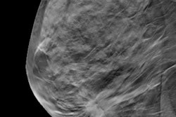
Mammography reduces deaths from breast cancer, but it also exposes patients to radiation and requires tissue compression, which some women find uncomfortable, wrote the team led by Lihong Wang, PhD. In addition, the technology's performance is compromised in dense breast tissue. Other breast imaging modalities, such as MRI, may involve contrast agents for optimum performance, Wang said in a statement released by the institute.
In an attempt to develop a less invasive, more effective breast cancer screening tool, Wang and colleagues built a scanner based on what they call breast photoacoustic CT (PACT), which uses the photoacoustic effect to convert absorbed optical energy into acoustic energy. PACT is capable of generating high-resolution images with deeper penetration than conventional optical imaging systems.
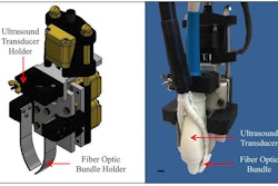
The group has developed a PACT scanner that features a table on which a woman lies prone with her breast in a recess containing ultrasound sensors and a laser. The device shines a near-infrared laser pulse into the breast tissue. The pulse spreads through the breast and is absorbed by hemoglobin molecules in the patient's red blood cells, causing the molecules to vibrate ultrasonically, according to the researchers.
The vibrations are tracked by an array of 512 ultrasonic sensors, the data from which are used to create an image of the breast's internal structures in a process that is similar to ultrasound imaging, the group wrote. PACT can image structures as small as 0.25 mm to a depth of 4 cm. The scan takes only 15 seconds -- in sharp contrast to other imaging modalities, such as breast MRI, which can take 45 minutes.
Wang and colleagues have scanned eight women using the PACT technology. The PACT exams identified eight out of nine tumors present in the pilot study participants, they found.
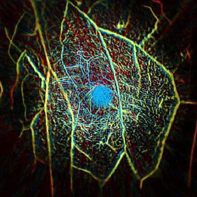
A false-color image that shows the vascular structure of a healthy human breast. Image courtesy of the California Institute of Technology.Wang has founded a company that licensed the PACT technology and will conduct large-scale clinical studies, he said. Going forward, he and his colleagues also plan to investigate the PACT technology for other uses, such as for assessing vascular tissue health in patients with diabetes.
"Our goal is to build a dream machine for breast screening, diagnosis, monitoring, and prognosis without any harm to the patient," he said. "We want it to be fast, painless, safe, and inexpensive."



![Examples of ultrasound findings and techniques. (A) Images in a 39-year-old male patient with a mass in the left thigh. The mass is heterogeneous on the B-mode US image (compared with the patient in D) and showed increased microvascularity (superb microvascular imaging [SMI]) and shear-wave elastography (SWE) values. Undifferentiated pleomorphic sarcoma was diagnosed at biopsy (with pleomorphic rhabdomyosarcoma in surgical specimen). (B) Images in an 18-year-old male patient with a mass in the left leg. The mass is hypoechoic on the B-mode image, with no other findings suggestive of malignancy. The lesion is in contact with the cortex of the tibia, which is slightly irregular. CT revealed a doubtful anteromedial tibial erosion. The microvascular study demonstrated high vascularization, suggestive of malignancy. Periosteal Ewing sarcoma was diagnosed with both histologic and immunohistochemical confirmation. (C) Images in a 69-year-old female patient with a lump growing on the outside of the left leg. Multiple SWE examinations were performed (please note the high values obtained in the measurements, whereas the color map highlights the stiffness relative to adjacent tissues). SMI showed areas of increased vascularization to target for sampling. Undifferentiated spindle cell sarcoma was diagnosed at biopsy, with residual leiomyosarcoma in the surgical specimen after neoadjuvant therapy. (D) Images in a 56-year-old female patient with a mass in the right thigh. The mass is heterogeneous at both B-mode ultrasound (similar to patient A) and MRI (coronal T2-weighted spectral attenuated inversion recovery [SPAIR]; T1-weighted pre-contrast and postcontrast imaging), which even shows uptake after the administration of paramagnetic contrast material, which is traditionally suggestive of malignancy. Low values at SMI and elastography are suggestive of benignity. Spindle cell lipoma was diagnosed at biopsy, with atypical spindle cell lipomatous tumor in the surgical specimen.](https://img.auntminnie.com/mindful/smg/workspaces/default/uploads/2026/08/images-radiol250278fig2.APCFLSvX6p.jpg?auto=format%2Ccompress&fit=crop&h=100&q=70&w=100)


![Examples of ultrasound findings and techniques. (A) Images in a 39-year-old male patient with a mass in the left thigh. The mass is heterogeneous on the B-mode US image (compared with the patient in D) and showed increased microvascularity (superb microvascular imaging [SMI]) and shear-wave elastography (SWE) values. Undifferentiated pleomorphic sarcoma was diagnosed at biopsy (with pleomorphic rhabdomyosarcoma in surgical specimen). (B) Images in an 18-year-old male patient with a mass in the left leg. The mass is hypoechoic on the B-mode image, with no other findings suggestive of malignancy. The lesion is in contact with the cortex of the tibia, which is slightly irregular. CT revealed a doubtful anteromedial tibial erosion. The microvascular study demonstrated high vascularization, suggestive of malignancy. Periosteal Ewing sarcoma was diagnosed with both histologic and immunohistochemical confirmation. (C) Images in a 69-year-old female patient with a lump growing on the outside of the left leg. Multiple SWE examinations were performed (please note the high values obtained in the measurements, whereas the color map highlights the stiffness relative to adjacent tissues). SMI showed areas of increased vascularization to target for sampling. Undifferentiated spindle cell sarcoma was diagnosed at biopsy, with residual leiomyosarcoma in the surgical specimen after neoadjuvant therapy. (D) Images in a 56-year-old female patient with a mass in the right thigh. The mass is heterogeneous at both B-mode ultrasound (similar to patient A) and MRI (coronal T2-weighted spectral attenuated inversion recovery [SPAIR]; T1-weighted pre-contrast and postcontrast imaging), which even shows uptake after the administration of paramagnetic contrast material, which is traditionally suggestive of malignancy. Low values at SMI and elastography are suggestive of benignity. Spindle cell lipoma was diagnosed at biopsy, with atypical spindle cell lipomatous tumor in the surgical specimen.](https://img.auntminnie.com/mindful/smg/workspaces/default/uploads/2026/08/images-radiol250278fig2.APCFLSvX6p.jpg?auto=format%2Ccompress&fit=crop&h=112&q=70&w=112)