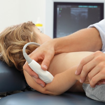
Ultrasound-guided radial artery cannulation in children is twice as successful as the traditional technique that has long been used to place cannulas, involving palpation of a patient's radial pulse, according to research published in the December issue of the Journal of Ultrasound in Medicine.
In addition, ultrasound for this application can be mastered even by relatively inexperienced clinicians, according to a team led by Dr. Nattachai Anantasit of Ramathibodi Hospital in Bangkok.
"Ultrasound-guided central venous access can improve the success rate, decrease complications, and reduce [healthcare] cost in critically ill ... children," the researchers wrote. "Moreover, ultrasound-guided vascular access skill can be acquired easily by a novice trainee."
Small vessels
Arterial catheterization is crucial for monitoring blood pressure and taking blood samples in pediatric patients. However, infants and very young children have small vessels, which tends to make artery cannulation difficult. Multiple attempts can cause complications such as artery spasms and bruising -- or even more serious ones such as hand ischemia, hemorrhage, or thrombosis, the researchers wrote.
That's why improving the first-time success rate of the procedure is so important, according to Anantasit and colleagues. Traditionally, radial artery cannulation has been done by palpating the patient's radial pulse, a technique that requires an experienced physician.
The use of ultrasound to guide arterial catheterization has been shown to be more effective than the palpation technique in adult populations, but there is less research that demonstrates whether it can also be used in children (J Ultrasound Med, December 2017, Vol. 36:12, pp. 2495-2501).
For the study, Anantasit and colleagues compared the success rate between the two techniques in a cohort of 84 critically ill pediatric patients between the ages of 1 month and 15 years who were admitted to a pediatric intensive care unit (median age, 22 months). Of these 84 patients, 43 were randomized to the palpation technique and 41 to the ultrasound-guided technique using a vascular transducer. The researchers sought to improve the success rate of radial artery cannulation from 40% to 75%.
The team defined procedure start time as when the needle initially penetrated the skin. Time to successful cannulation was measured until the removal of the needle, and success was defined as cannulation of the radial artery in three or fewer attempts. Staff members who performed the procedure consisted of four first-year and three second-year pediatric critical care fellows; all had experience of more than 10 cases in either the ultrasound-guided or palpation technique.
Anantasit and colleagues found that total success and first-attempt rates for the ultrasound-guided group were significantly higher than for the palpation group -- and median time to successful cannula placement and hematoma incidence were significantly lower. There were no major complications in either group.
| Palpation vs. ultrasound-guided arterial cannulation in children | ||
| Palpation group | Ultrasound group | |
| Total success rate | 39.5% | 80.5% |
| 1st-attempt success rate | 29.4% | 60.6% |
| Median time to cannulation | 10.4 minutes | 3.3 minutes |
| Incidence of hematoma | 53.3% | 12.5% |
"Critically ill children were found to benefit from ultrasound-guided radial artery cannulation in terms of higher first-attempt success rate, shorter time for cannulation, and fewer complications," the group wrote.
Excellent alternative
The study findings suggest that ultrasound offers an excellent alternative to palpation for radial artery cannulation in children -- and that it can be handled well even by less-experienced clinicians, Anantasit and colleagues wrote.
"We recommend ultrasound-guided radial artery cannulation in [this population, and] suggest that young physicians should practice the traditional palpation technique first, learn the ultrasound-guided technique to help in difficult cases, and practice both techniques to maintain their skills," the group concluded.


![Examples of ultrasound findings and techniques. (A) Images in a 39-year-old male patient with a mass in the left thigh. The mass is heterogeneous on the B-mode US image (compared with the patient in D) and showed increased microvascularity (superb microvascular imaging [SMI]) and shear-wave elastography (SWE) values. Undifferentiated pleomorphic sarcoma was diagnosed at biopsy (with pleomorphic rhabdomyosarcoma in surgical specimen). (B) Images in an 18-year-old male patient with a mass in the left leg. The mass is hypoechoic on the B-mode image, with no other findings suggestive of malignancy. The lesion is in contact with the cortex of the tibia, which is slightly irregular. CT revealed a doubtful anteromedial tibial erosion. The microvascular study demonstrated high vascularization, suggestive of malignancy. Periosteal Ewing sarcoma was diagnosed with both histologic and immunohistochemical confirmation. (C) Images in a 69-year-old female patient with a lump growing on the outside of the left leg. Multiple SWE examinations were performed (please note the high values obtained in the measurements, whereas the color map highlights the stiffness relative to adjacent tissues). SMI showed areas of increased vascularization to target for sampling. Undifferentiated spindle cell sarcoma was diagnosed at biopsy, with residual leiomyosarcoma in the surgical specimen after neoadjuvant therapy. (D) Images in a 56-year-old female patient with a mass in the right thigh. The mass is heterogeneous at both B-mode ultrasound (similar to patient A) and MRI (coronal T2-weighted spectral attenuated inversion recovery [SPAIR]; T1-weighted pre-contrast and postcontrast imaging), which even shows uptake after the administration of paramagnetic contrast material, which is traditionally suggestive of malignancy. Low values at SMI and elastography are suggestive of benignity. Spindle cell lipoma was diagnosed at biopsy, with atypical spindle cell lipomatous tumor in the surgical specimen.](https://img.auntminnie.com/mindful/smg/workspaces/default/uploads/2026/08/images-radiol250278fig2.APCFLSvX6p.jpg?auto=format%2Ccompress&fit=crop&h=100&q=70&w=100)


![Examples of ultrasound findings and techniques. (A) Images in a 39-year-old male patient with a mass in the left thigh. The mass is heterogeneous on the B-mode US image (compared with the patient in D) and showed increased microvascularity (superb microvascular imaging [SMI]) and shear-wave elastography (SWE) values. Undifferentiated pleomorphic sarcoma was diagnosed at biopsy (with pleomorphic rhabdomyosarcoma in surgical specimen). (B) Images in an 18-year-old male patient with a mass in the left leg. The mass is hypoechoic on the B-mode image, with no other findings suggestive of malignancy. The lesion is in contact with the cortex of the tibia, which is slightly irregular. CT revealed a doubtful anteromedial tibial erosion. The microvascular study demonstrated high vascularization, suggestive of malignancy. Periosteal Ewing sarcoma was diagnosed with both histologic and immunohistochemical confirmation. (C) Images in a 69-year-old female patient with a lump growing on the outside of the left leg. Multiple SWE examinations were performed (please note the high values obtained in the measurements, whereas the color map highlights the stiffness relative to adjacent tissues). SMI showed areas of increased vascularization to target for sampling. Undifferentiated spindle cell sarcoma was diagnosed at biopsy, with residual leiomyosarcoma in the surgical specimen after neoadjuvant therapy. (D) Images in a 56-year-old female patient with a mass in the right thigh. The mass is heterogeneous at both B-mode ultrasound (similar to patient A) and MRI (coronal T2-weighted spectral attenuated inversion recovery [SPAIR]; T1-weighted pre-contrast and postcontrast imaging), which even shows uptake after the administration of paramagnetic contrast material, which is traditionally suggestive of malignancy. Low values at SMI and elastography are suggestive of benignity. Spindle cell lipoma was diagnosed at biopsy, with atypical spindle cell lipomatous tumor in the surgical specimen.](https://img.auntminnie.com/mindful/smg/workspaces/default/uploads/2026/08/images-radiol250278fig2.APCFLSvX6p.jpg?auto=format%2Ccompress&fit=crop&h=112&q=70&w=112)