Is contrast-enhanced ultrasound (CEUS) effective for differentiating between inflammatory bowel disease (IBD) and colon cancer? The answer appears to be yes, but more research is needed, according to a study published online November 7 in Ultrasound in Medicine and Biology.
Transabdominal ultrasound is increasingly being used to diagnose intestinal diseases, as it is a noninvasive, convenient imaging modality that visualizes the thickness of the intestinal walls, the extent and blood flow of lesions, and the shape of the intestinal cavity, wrote researchers from Peking University Third Hospital in Beijing. However, it can be difficult for 2D ultrasound to differentiate between two common intestinal diseases, IBD and colon cancer, which have similar clinical features. That's why CEUS may be more effective.
"Although both inflammation and tumors manifest the characteristic of angiogenesis, they are significantly different in blood perfusion, which is helpful in differential diagnosis between the two diseases," lead author Dr. Fan Zhang and colleagues wrote. "CEUS is a new technology for evaluation of microvessels and tissue perfusion. Our study aimed to evaluate the diagnostic performance of CEUS in the differential diagnosis of IBD and colon cancer."
Noninvasive alternative
Typically, colonoscopy and gastrointestinal x-ray are used to diagnose gastrointestinal tract diseases because they offer clear imaging of the mucosal surface. But these procedures can't image submucosal lesions as well as clinicians would like, the authors wrote.
Ultrasound can visualize the degree of abdominal wall thickening, internal stratification and structure, and the extent and depth of the lesion and lymph node metastasis, but it hasn't been used for gastrointestinal scans because of limitations such as gas interference, operator dependence, and low sensitivity in displaying blood flow (Ultrasound Med Biol, November 7, 2017).
CEUS addresses some of those limitations, according to the group.
"CEUS may cover some of the shortages of ultrasound by displaying the blood perfusion in intestinal lesions in detail, which could accurately reflect the perfusion difference between inflammation and tumors, without the disadvantages of enhanced CT and MRI, such as high cost, radioactivity, and toxicity to the kidney," the authors wrote.
For their study, Zhang and colleagues included 51 patients with thickened bowel walls. Of these, 13 had IBD and 38 had colon cancer. All patients underwent a conventional ultrasound exam and a contrast-enhanced exam. Two radiologists reviewed and analyzed each patient's medical records and imaging exam results and came to a consensus on the features of ultrasound and CEUS parameters, according to Zhang's group.
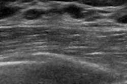
On unenhanced ultrasound, the researchers found that characteristics of IBD included swelling of the mucosal to submucosal layers (69.2%) or all layers of the intestinal wall (30.8%), with a mean intestinal wall thickness of 0.71 cm. In the tumor group, the intestinal walls showed heterogeneous thickening of the whole wall, with a mean thickness of 1.22 cm. Intestinal wall stratification was preserved in 69.2% of the IBD group but only 2.6% of the tumor group.
On CEUS, 12 of the 13 cases of IBD showed "ordered" enhancement (i.e., consistent visualization of "comb-teeth-like" vessels that represent arterial branching within the intestinal wall). But in the tumor group, disordered enhancement was found in 94.7% of cases, the researchers wrote.
Better diagnosis?
This difference in how the two diseases show enhancement on contrast-enhanced ultrasound is related to their pathologic characteristics and can help distinguish between them, Zhang and colleagues noted.
"The features of IBD are preserved stratification of the thickened intestinal wall on ultrasound and 'comb-teeth-like' vessels ... on CEUS," they wrote. "Colon cancer was totally different from IBD, featuring loss of intestinal wall stratification on ultrasound and disordered ... enhancement on CEUS."
The study results are promising, but more research is needed, Zhang and colleagues wrote.
"CEUS may prove useful for the differential diagnosis of IBD and colon cancer, but more studies are required," they concluded.