An image processing technique applied to contrast-enhanced ultrasound can deliver sharp, spatially compounded images without the need for additional transmit and receive cycles that can damage microbubble contrast, researchers have concluded.
In a presentation at ECR 2017 in Vienna, ultrasound executive Glen McLaughlin, PhD, showed how spatial compounding enhances image quality in microbubble contrast-enhanced ultrasound by "recycling" acoustic echo information from the ultrasound transducer multiple times, extracting more image information from each cycle.
In a comparison with standard imaging techniques, the so-called synthetic steered continuous transmit/receive focusing (SSCTRF) images had better image quality and higher contrast-to-noise ratios and improved diagnostic confidence.
"Being able to extract more information from the images that are coming out allows you to get an additional dimension of time within your images, reducing those constraints to derive better diagnostic performance," said McLaughlin, worldwide chief product officer at ultrasound developer Mindray.
McLaughlin and colleagues David Napolitano, Robert Steins, and Jim Baun from the Mindray Innovation Center in Mountain View, CA, applied the SSCTRF technique to obtain spatially compounded images from an ultrasound microbubble contrast sequence without additional transmit or receive cycles, comparing them with standard B-mode images.
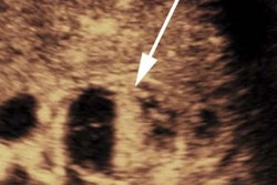
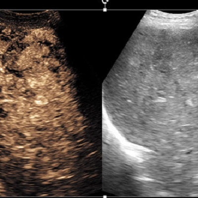
 Contrast wash-in demonstrates the ability of contrast agents to enhance the visualization of tumors. Amber image (left) is the contrast image (cubic fundamental with synthetic spatial compounding); image on right is simple fundamental B-mode image. Images are of the liver, using a C4-1 transducer. All images courtesy of Glen McLaughlin, PhD.
Reusing ultrasound data
By recycling channel domain data, additional information can be extracted from the signal, McLaughlin explained.
"What is channel recycling? It's the ability to process the echoes from each transmit/receive cycle multiple times, and in doing so being able to extract images which can reduce the overall coherence of the correlation between the two frames and achieve spatially compounded images," he said.
Multiple signals can be extracted from a single transmit sequence, including fundamental, harmonic, and third harmonic images. Domain data recycling offers the ability to steer each frame in a total of three directions -- left, right, and straight ahead, for example -- delivering 12 total images from four transmit/receive cycles, he said.
"So why do we care?" McLaughlin asked. "As practitioners, we know that ultrasound is an image mode of trade-offs. There is no one ideal path. You're either getting very good temporal resolution, very good spatial resolution, or very good penetration, but it's really hard to get all of them working well simultaneously. Because time is not your friend."
 Synthesized spatially compounded image (above) of the thyroid versus original image (below) shows a reduction in speckle spatial variance and higher contrast resolution.
Image faceoff
To compare traditional images with the enhanced SSCTRF images, the researchers collected contrast ultrasound loops from a Zonare ZS3 ultrasound system (Mindray) using two different transducers. The loops were processed with traditional methods, extracting single cubic-fundamental and single B-mode images.
The image data were also processed to extract up to six cubic-fundamental images and three harmonic images along with three B-mode images. The six cubic-fundamental images consisted of two images created by weighting and adding combinations of the pulse sequence, and four images that were then constructed through SSCTRF techniques to generate two steered-left and two steered-right images. The same technique was used to generate three standard harmonic and three B-mode images. The traditional images were compared with the SSCTRF images.
Image loops generated using SSCTRF techniques showed improved contrast resolution, better signal-to-noise ratios, and sharper border delineation, and preserved tissue perfusion kinetics without destroying additional microbubbles, providing enhanced visualization and higher diagnostic confidence, McLaughlin said.
The technique can "effectively compress the point-spread function to get an effective transmit focus at every point throughout the [field-of-view] while not requiring a true physical focus," he said.
 Spatial coherence factor comparison between compounding views.
Extracting more information from the signal adds the element of time to the image data, reducing ultrasound's inherent constraints and driving better diagnostic performance, he concluded.
Trade-offs are especially steep with the use of microbubble contrast, because bubbles break during every transmit/receive cycle, so it's important to minimize them while maximizing the information from each cycle, McLaughlin said. Toward that end, channel data recycling with SSCTRF techniques is a viable path toward improving signal-to-noise ratio and contrast resolution while maintaining temporal resolution and avoiding additional bubble destruction.