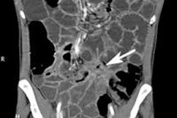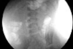
There's no need to use abdominal radiography to diagnose pediatric intussusception -- ultrasound is better, and it does not expose children to radiation, according to a new study published in the journal Pediatric Surgery International.
The disorder, which is the leading cause of bowel obstruction in infants and preschoolers, occurs when part of the intestine slides into an adjacent section, according to researchers from Ireland. Classic symptoms include abdominal pain, "red currant jelly" stools, and a palpable mass, but these are only present in about one-third of cases.
Because the disorder occurs primarily in nonverbal patients, clinicians must use imaging to make a quick and accurate diagnosis. Failure to identify the disorder can lead to bowel ischemia, perforation, peritonitis, shock, and even death.
No need for x-ray
Abdominal x-ray followed by a contrast enema has been the traditional diagnostic tool for the disorder, wrote Dr. Farhan Tareen and colleagues from Our Lady's Children's Hospital, Crumlin in Dublin. But over the past three decades, ultrasound has been shown to perform better than abdominal x-ray, with a sensitivity rate of nearly 100% and a false-negative rate of less than 1%.
Contrast enema has been replaced by a procedure called pneumatic reduction of intussusception (PRI), during which air is pumped into the area so that the intestine can be placed back into position. PRI has a success rate of up to 90% (Pediatr Surg Int, November 6, 2015).
Tareen's group investigated the performance of abdominal radiography in diagnosing intussusception, predicting the outcome of PRI, and identifying the presence of air or gas in the abdominal cavity (pneumoperitoneum).
To do this, the researchers analyzed 651 cases of intussusception that had been treated at two pediatric surgery centers between 1998 and 2013. The age range of the patients was 3 days to 13 years, with a median age of 7 months; the four most common presenting symptoms were irritability/abdominal pain (84%), vomiting (81%), bleeding from the rectum (43%), and palpable abdominal mass (45%).
Of the 651 cases, 644 were treated with PRI, and all of these patients underwent diagnostic ultrasound before the procedure was performed. PRI was successful 85.4% of the time; in 13.2% of cases, it failed to reduce the intussusception, and in 1.4% of cases, it resulted in perforation of the bowel. In 67 cases, or 10.4%, there was a recurrence of the intussusception.
Of the patients treated with PRI, 412 had an abdominal x-ray before the procedure; 74% had findings that suggested intussusception, while 26% of the exams were normal. But ultrasound was also performed to confirm the diagnosis -- which suggests that abdominal x-ray was not considered adequate for diagnosis, according to Tareen and colleagues.
"Twenty-six percent of radiographs in cases of confirmed intussusception were normal in this large series," the researchers wrote. "[The use of abdominal radiography] for diagnostic purposes is not supported by our findings or the wider literature."
In addition, positive findings on abdominal radiography did not suggest poorer PRI outcomes, compared with patients who had negative abdominal x-rays. Also, PRI outcomes in children who did not have an abdominal x-ray beforehand did not differ significantly from those who did. Finally, radiography did not identify pneumoperitoneum in any of the patients.
What's the bottom line? Abdominal radiography is not recommended for diagnosing intussusception in children, the researchers concluded. Rather, doctors should prioritize ultrasound and specialist assessment.
"[Abdominal radiography] is inaccurate in the diagnosis of intussusception, does not correlate with the outcome of PRI, and has not been shown to detect occult pneumoperitoneum," the group wrote.


![Examples of ultrasound findings and techniques. (A) Images in a 39-year-old male patient with a mass in the left thigh. The mass is heterogeneous on the B-mode US image (compared with the patient in D) and showed increased microvascularity (superb microvascular imaging [SMI]) and shear-wave elastography (SWE) values. Undifferentiated pleomorphic sarcoma was diagnosed at biopsy (with pleomorphic rhabdomyosarcoma in surgical specimen). (B) Images in an 18-year-old male patient with a mass in the left leg. The mass is hypoechoic on the B-mode image, with no other findings suggestive of malignancy. The lesion is in contact with the cortex of the tibia, which is slightly irregular. CT revealed a doubtful anteromedial tibial erosion. The microvascular study demonstrated high vascularization, suggestive of malignancy. Periosteal Ewing sarcoma was diagnosed with both histologic and immunohistochemical confirmation. (C) Images in a 69-year-old female patient with a lump growing on the outside of the left leg. Multiple SWE examinations were performed (please note the high values obtained in the measurements, whereas the color map highlights the stiffness relative to adjacent tissues). SMI showed areas of increased vascularization to target for sampling. Undifferentiated spindle cell sarcoma was diagnosed at biopsy, with residual leiomyosarcoma in the surgical specimen after neoadjuvant therapy. (D) Images in a 56-year-old female patient with a mass in the right thigh. The mass is heterogeneous at both B-mode ultrasound (similar to patient A) and MRI (coronal T2-weighted spectral attenuated inversion recovery [SPAIR]; T1-weighted pre-contrast and postcontrast imaging), which even shows uptake after the administration of paramagnetic contrast material, which is traditionally suggestive of malignancy. Low values at SMI and elastography are suggestive of benignity. Spindle cell lipoma was diagnosed at biopsy, with atypical spindle cell lipomatous tumor in the surgical specimen.](https://img.auntminnie.com/mindful/smg/workspaces/default/uploads/2026/08/images-radiol250278fig2.APCFLSvX6p.jpg?auto=format%2Ccompress&fit=crop&h=100&q=70&w=100)


![Examples of ultrasound findings and techniques. (A) Images in a 39-year-old male patient with a mass in the left thigh. The mass is heterogeneous on the B-mode US image (compared with the patient in D) and showed increased microvascularity (superb microvascular imaging [SMI]) and shear-wave elastography (SWE) values. Undifferentiated pleomorphic sarcoma was diagnosed at biopsy (with pleomorphic rhabdomyosarcoma in surgical specimen). (B) Images in an 18-year-old male patient with a mass in the left leg. The mass is hypoechoic on the B-mode image, with no other findings suggestive of malignancy. The lesion is in contact with the cortex of the tibia, which is slightly irregular. CT revealed a doubtful anteromedial tibial erosion. The microvascular study demonstrated high vascularization, suggestive of malignancy. Periosteal Ewing sarcoma was diagnosed with both histologic and immunohistochemical confirmation. (C) Images in a 69-year-old female patient with a lump growing on the outside of the left leg. Multiple SWE examinations were performed (please note the high values obtained in the measurements, whereas the color map highlights the stiffness relative to adjacent tissues). SMI showed areas of increased vascularization to target for sampling. Undifferentiated spindle cell sarcoma was diagnosed at biopsy, with residual leiomyosarcoma in the surgical specimen after neoadjuvant therapy. (D) Images in a 56-year-old female patient with a mass in the right thigh. The mass is heterogeneous at both B-mode ultrasound (similar to patient A) and MRI (coronal T2-weighted spectral attenuated inversion recovery [SPAIR]; T1-weighted pre-contrast and postcontrast imaging), which even shows uptake after the administration of paramagnetic contrast material, which is traditionally suggestive of malignancy. Low values at SMI and elastography are suggestive of benignity. Spindle cell lipoma was diagnosed at biopsy, with atypical spindle cell lipomatous tumor in the surgical specimen.](https://img.auntminnie.com/mindful/smg/workspaces/default/uploads/2026/08/images-radiol250278fig2.APCFLSvX6p.jpg?auto=format%2Ccompress&fit=crop&h=112&q=70&w=112)