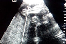
It was a heartbreaking story, but it could have been even more tragic were it not for the Mobile Ultrasound Patrol in Morocco. After being given an ultrasound in her rural village, a young woman in pain -- unaware that she was pregnant -- was found to be carrying two dead fetuses.
Based on the ultrasound findings and the gestational age of the fetuses, the woman was transported to a nearby hospital and given lifesaving treatment. That story is one of many successes from the Mobile Ultrasound Patrol, a project that brought together vendors, physicians, and nurses to facilitate onsite ultrasound studies for pregnant women at three rural Moroccan villages in early 2014.
From the nearly 600 exams performed during the study, 94 high-risk pregnancies were diagnosed, according to image sharing software developer Trice Imaging, which initiated the project and provided the mobile encryption and image management software. The studies were transmitted wirelessly over 3G networks for remote second opinions from physicians based in Rabat and Casablanca in Morocco and also in Paris.
Maternal care gap
In Morocco, 800 women die every day from maternal complications due largely to geographical distance from good healthcare and the low number of physicians; there are approximately 60 doctors per 100,000 in the population, said Johanna Wollert Melin, chief operating officer and co-founder of Trice.
There's also a lack of equipment, including ultrasound, or any type of prenatal care. Furthermore, many of the country's healthcare workers don't have the necessary education.
"And we know that this is the case with a lot of African countries," she said.
Approximately 2% to 3% of women in rural parts of Africa are dying because they don't receive adequate care, according to Melin.
The Mobile Ultrasound Patrol project got underway after Trice applied for funds from wireless firm Qualcomm's Wireless Reach program, which aims to bring wireless technologies to underserved communities around the world. In addition to the Qualcomm funding, three M-Turbo compact ultrasound devices were provided by Fujifilm SonoSite.
How it works
Image transmission was relatively straightforward. After the images were acquired, a wireless dongle transmitted the images over Wi-Fi to a Sony smartphone or tablet. The Trice application then encrypted the images and sent them via a 3G mobile network to the Trice cloud-based image management platform.
From there, experienced physicians based in Rabat, Casablanca, and Paris -- alerted to the exam by an encrypted email -- could log on to the system to provide second opinions on the studies. The scans were performed in three rural villages: Oulmes, Boulemane, and Ribat el Kheir.
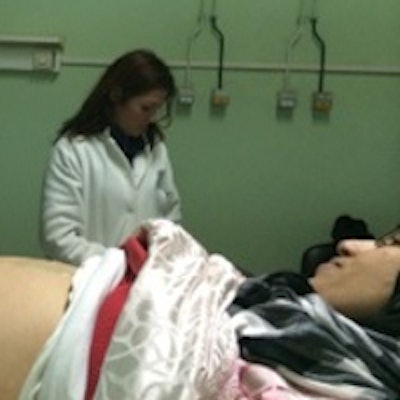
 A pregnant woman receives a prenatal ultrasound exam as part of the Mobile Ultrasound Patrol initiative. Image courtesy of Trice Imaging.
Before the technology could be deployed, a human network needed to be assembled, said Dagmar Nuber, Trice's business development manager and onsite leader for the project.
"Even if you have the local people doing the scans, you need a network of doctors reviewing the scans," Nuber said.
After signing on the experienced doctors in Morocco and France to provide second opinions, project organizers held a training session in December 2013 for those who would be scanning patients in the field. Sonographers were trained on the scanning protocol, they performed live scans on pregnant women, and they were then certified by a physician to be part of the trial, which took place for four weeks beginning in January 2014.
"The mission of the Mobile Ultrasound Patrol project was to validate our wireless technology and connect the portable ultrasound devices to provide access to state-of-the-art imaging diagnostic tools in places it's never been before, to prove that you can actually do diagnosis, have high image quality, and lower the cost and actually shorten the time [to diagnosis]," Melin said.
After the four-week trial, participants returned on their own every second week to the villages for follow-up visits, Nuber said. Overall, 575 exams were performed in health houses at the three villages.
The project wirelessly transferred 3,108 images, 92% of which could be used for professional diagnosis, according to Trice. Thanks to the Mobile Ultrasound Patrol, the average time to get a second opinion on a patient was reduced from two weeks to under 24 hours, and the average time to transport medical data dropped from two weeks to one minute, the company said. The cost for a second opinion fell from $80 per patient to $2.
While physicians -- either generalists or ob/gyns -- would perform scans onsite for diagnosis, midwives from the health houses also performed a few limited scans each day, Nuber said.
"They are not allowed to do a complete scan," she said. "It's just to be assured that this delivery can be done in the health house or if it has to be done somewhere else."
Physicians involved in the project reported many positive benefits, including discovering many high-risk pregnancies, finding an early case of uterine cancer, and identifying pregnancies that would be difficult deliveries, Nuber said.
Current status
The formal trial has concluded, but ultrasound studies are still being performed in Morocco.
"Once or twice a month, they are in those regions," Nuber said. "They go out to different health houses."
The Moroccan government is now considering whether to buy the portable ultrasound devices together with the Trice software, Melin said. Two of the regional health governments have elected to move forward with the project.