As we discussed in part 1 of our two-part series on tennis elbow, you can develop this painful condition without playing tennis or racket sports. Repetitive movements on the job, such as the daily work of a sonographer, can also trigger tennis elbow.
In the first article, we shared some simple stretching exercises to improve mobility and flexibility to relieve elbow pain. Now we'll add three simple exercises to improve stability and strength. You should train two or three days a week, and pick one stretching exercise and one strengthening exercise to perform each day that you work out.
Strength and stability
Wrist curls
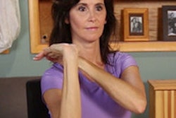
First, hold some light-resistance weights in your hands. Be sure to use a weight that is easy for you to handle. Begin by sitting on the edge of a stable chair or secure bench. Then rest your forearms on your thighs with your palms facing inward and your thumbs up (figure 1).
Now bend the wrists up toward the ceiling using as much range of motion as possible. Think about keeping your thumbs up and pushing them back toward your body (figure 2).
 Figures 1 (left) and 2 (right). All images courtesy of Mark Roozen and Doug Wuebben.
Next, lower the weights in your hands down past your knees, working to get the little finger and outside of your hand to touch below your knee cap. Keep your elbows and forearms on your thighs during the whole movement. It's important not to get movement by lifting the elbow off the thigh.
Change your position each time you do the wrist curl exercise; each hand position will isolate or work different muscles in the forearms and wrist. The palms-up position is shown in figures 3 and 4.
 Figures 3 (left) and 4 (right).
The palms-down position is demonstrated in figures 5 and 6.
 Figures 5 (left) and 6 (right).
Stability ball walk out
The stability ball walk out develops grip hand strength, forearm strength, and shoulder and chest strength, along with stability in the core.
First, lie on top of a stability ball, with your hands on the ground and knees against the ball for support and control. Then begin to walk your hands out away from the ball, while at the same time extending (straightening) your legs (figure 7).
Walk out as far as possible, while still being able to control balance and stability. Progress from the hips to the thighs, knees, shins, feet, and toes (figure 8).
 Figures 7 (left) and 8 (right).
To make the exercise more difficult, keep your feet closer together and work your way farther out from the ball. This will increase the need for stability through the arms, shoulders, chest, and core. Either way, once you've reached the maximum walk-out point for you, walk your hands back toward the ball and repeat for a set number of reps.
Miniball/Medicine ball wall pushup
The ball wall pushup works the hands, forearms, arms, shoulders, and chest and can be a great core exercise for stability. By changing hand position and the position of the ball, you can also isolate, or work, different muscles in the forearm and wrist.
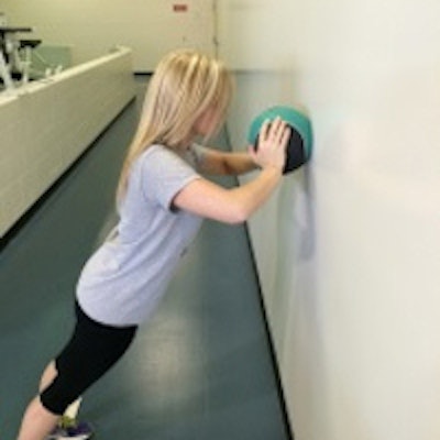
Begin by holding a small ball, a medicine ball, or even a stability ball lightly in your hands. Stand upright and hold the ball against the wall. Keep your body in a straight line. The ball should initially be at shoulder and chest level (figure 9).
Maintaining good balance (the wider your feet, the more stable you will be), extend (straighten) your arms while holding the ball against the wall. Then flex, or bend, the arms at the elbows to bring your chest in to touch the ball. If you cannot bring your chest all the way to the ball, use as much range of motion as possible (figure 10).
 Figures 9 (left), 10 (center), and 11 (right).
Keep in mind that the smaller the ball, the harder the exercise will be. So you may want to start with a stability ball, beach ball, or basketball/volleyball, etc. Once you feel comfortable with the exercise, you can also change the position of the ball, such as placing it at head or stomach level (figure 11).
Note that it's very important not just to move your head toward the ball or wall. The arms must bend and straighten to benefit from this exercise.
Doug Wuebben is a registered echocardiographer and also a consultant, national presenter, and author of e-books in the areas of ergonomics, exercise and pain, and injury correction for sonographers. He has also been published on the topics of telemedicine and achieving lab accreditation, and he can be reached at [email protected]. Mark Roozen is a strength coach with the Cleveland Browns and also the owner and president of Performance Edge Training Systems (PETS).
The comments and observations expressed herein are those of the authors and do not necessarily reflect the opinions of AuntMinnie.com.