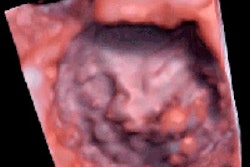
A joint committee of the European Association for Echocardiography and the American Society of Echocardiography has issued the first joint guidelines for image acquisition using 3D echocardiography.
The recommendations, published in the Journal of the American Society of Echocardiography (JASE, January 2012, Vol. 25:1, pp. 3-46), are a practical guide for acquiring, analyzing, and displaying various cardiac structures using 3D echo. Dr. Roberto Lang from Mount Sinai Medical Center in New York City and colleagues discuss the advantages and limitations of the technique, as well as current and potential uses of the technology, exploring novel image acquisition and display techniques.
The paper details data acquisition modes ranging from simultaneous multiplane mode to real-time 3D mode, as well as narrow to focused wide-sector zoom. The authors also discuss challenges of 3D acquisition, including 3D gating and breath-hold and 3D optimization tips.
In addition, the group discusses the problems associated with low- and high-gain settings, advising that practitioners begin in the midrange (50 units), then optimize settings with slightly higher time-gain compensation to enable the greatest flexibility in postprocessing.
Transthoracic and transesophageal exam techniques also are covered in detail. In transthoracic 3D echo, for example, the authors advise that clinicians acquire images from multiple transthoracic echo transducer positions to overcome the limitations of poor temporal and spatial resolution. For transesophageal exams, gated 3D modes, including 3D color-flow Doppler, also should be used whenever ECG and respiration gating requirements permit, in order to benefit from the improved spatial and temporal resolution of the wide-angled acquisitions, the authors stated.
Foremost among 3D echo's limitations are that spatial and temporal resolution remain poor, but this is expected to improve as the technology advances, they concluded. Live 3D echo color Doppler acquisition is limited to small color Doppler volumes, generally with limited temporal resolution of 10 to 15 voxels/sec.
"Alternatively, multiple-beam full-volume acquisition of color Doppler providing larger color Doppler volumes and volume rates (up to 40 voxels/sec) are limited by stitching artifacts, resulting in significant displacement between different subvolumes," Lang and colleagues wrote.
Although 3D echocardiography has been a research interest for more than three decades, recent improvements in imaging technology have made it suitable for clinical use, they concluded.
In a statement accompanying release of the article, Dr. Alan Pearlman, JASE editor in chief, wrote: "I believe that real-time volumetric imaging using 3D echocardiography now provides a practical way to measure ventricular volumes and ejection fraction without making any assumptions about chamber geometry, and suspect that one day this imaging method will surpass current 2D echo imaging, just as 25 years ago 2D imaging supplanted M-mode echocardiography."