A new echocardiography software package released this month analyzes cardiac structure and function pixel by pixel to render detailed functional 3D images that could speed up diagnosis and reduce the number of exams needed in patients undergoing cardiac workups.
GE Healthcare's cSound software, which received 510(k) U.S. Food and Drug Administration (FDA) clearance earlier this year, is being bundled into its Vivid series of advanced ultrasound systems (S70, E90, E95) for marketing in the U.S. and some countries in Europe, Asia, and Latin America, GE said. The first system is already in use at Aurora St. Luke's Medical Center in Milwaukee.
The software works by collecting large amounts of data from the patient's cardiovascular system and selecting precise, pixel-by-pixel information to use in generating images, GE said.
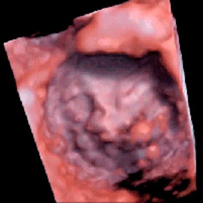
 3D view of the mitral valve in motion. Image courtesy of GE Healthcare.
Analyzing data in 1 pass
The problem with conventional hardware-based beamforming ultrasound scanners is that they're limited to processing one piece of data at a time, losing some of it in the process and resulting in unclear images, according to the company. In contrast, cSound stores large amounts of channel data, and it does this faster than conventional systems do.
Software-based beamforming identifies the best image data pixel by pixel and then converts the information into meaningful images of the heart. In conventional hardware-based beamforming, this is performed segment by segment and filled in layer by layer to eventually form an advanced image, GE said. However, in software-based beamforming, the software analyzes all of the data at the start of the process, producing a single, high-quality image.
How does the software determine the "best" data from the echo signal using all of the image data? It's based on a specific algorithm, adaptive contrast enhancement (ACE), which determines the quality of the channel data contributing to each pixel of the image, according to Erik Steen, GE's principal engineer for cardiovascular ultrasound.
"This is a large number of data samples, typically in the thousands," Steen wrote in an email to AuntMinnie.com.
ACE is based on statistical analysis of these data, and it works by estimating the likelihood of the data originating from real tissue versus clutter or image noise. It works equally well with normal and abnormal heart anatomy, he said.
Another cSound feature is a volume-rendering algorithm known as HDlive, which combines several methods to render the heart in real-time from echo data, according to Steen. Volume-rendering algorithms treat volume data as a set of semitransparent voxels. Surfaces are then defined implicitly by robust volume gradient calculations.
"Depth coloring, shadowing, and light reflections are combined to provide detailed and realistic-looking images with enhanced depth perception," Steen said.
HDlive doesn't rely on atlases or templates of any kind, but it does benefit from the improved volume image quality provided by cSound, he added.
The GE promotional video below shows software-rendered images of the heart in motion.
Transthoracic or transesophageal echo
In cardiac patients, cSound can be used transesophageally or transthoracically, depending on the application, according to Al Lojewski, GE's general manager of cardiovascular ultrasound.
"They'll typically be doing transesophageal during an intervention, repairing or replacing a valve; they'll also do that for extremely difficult patients," such as obese heart patients, Lojewski said in an interview with AuntMinnie.com.
However echocardiography is performed, the limitations with conventional echo systems are the same, he said: They simply lack sufficient anatomic and functional detail to make a confident diagnosis.
"It's been published that in 10%, 14%, up to 25% [of cases] we can't see the images because maybe the body size is too big, or we can't get between the ribs of a smaller, elderly lady," Lojewski said. "Or maybe it's difficult to see the valve placement, and it leads to more tests and more expense, or exams that involve radiation. With this technology, we believe that we have significantly reduced the number of patients on whom you can't do an exam."
Given recent advances in detailed visualization of cardiac function, ultrasound has the potential to rapidly transform how patients are cared for over the next decade, especially in cardiovascular care, according to Lojewski.
"We envision a day when all cardiologists can see inside any heart, providing more directed care for each patient," he said.
cSound's price point ranges from about $30,000 for a laptop-sized ultrasound machine up to about $250,000 depending on how the scanner is equipped, Lojewski said.