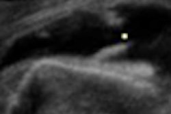
Certain ultrasound criteria can be useful in distinguishing between malignant and benign thyroid nodules, but their accuracy depends on the size of the tumor, according to a multicenter study published in the June issue of Radiology.
"Application of US criteria for the differentiation of malignant from benign nodules may help in accurate diagnosis of thyroid nodules," according to the Thyroid Study Group of the Korean Society of Neuro and Head and Neck Radiology.
The researchers sought to retrospectively evaluate the diagnostic accuracy of ultrasound criteria for the depiction of benign and malignant thyroid nodules, evaluating 8,024 consecutive patients who had undergone thyroid ultrasound at nine affiliated hospitals between January 2003 and June 2003. Of the 8,024 patients, 831 patients with 849 nodules (360 malignant) that were diagnosed at surgery or biopsy were included in the study (Radiology, June 2008, Vol. 247:3, pp. 762-770).
All ultrasound exams were performed using either an HDI 5000 (Philips Healthcare, Andover, MA) or Sequoia scanner (Siemens Healthcare, Erlangen, Germany) equipped with a linear-array transducer of 5-12 MHz or 8-15 MHz . Both transverse and longitudinal real-time imaging were performed.
Three radiologists retrospectively reviewed all the ultrasound images, evaluating nodule size, presence of spongiform appearance, shape, margin, echotexture, and echogenicity, as well as the presence of microcalcification, macrocalcification, or rim calcification.
The researchers found that taller-than-wide shape, a spiculated margin, marked hypoechogenicity, microcalcification, and macrocalcification were statistically significant findings for malignancy (p < 0.05).
|
Ultrasound findings for benign nodules were isoechogenicity (44.2% sensitivity, 90.8% specificity) and a spongiform appearance (10.4% sensitivity, 99.7% specificity). The researchers noted that the presence of at least one malignant ultrasound finding had a sensitivity of 83.3%, a specificity of 74%, and a diagnostic accuracy of 78%.
The sensitivity of microcalcifications was also lower in nodules with a diameter of less than 1 cm (36.6%) than in larger nodules (51.4%, p < 0.05).
"When US features suggestive of a benign nodule (an ovoid-to-round shape, a well-defined smooth margin, isoechogenicity, and a spongiform appearance) were compared, no significant difference in their frequency was found between malignant nodules smaller than 10 mm and those larger than 10 mm," the authors wrote. "In contrast, benign nodules larger than 10 mm had more isoechogenicity (62.6% versus 35.5%, p < 0.001) and a well-defined smooth margin (78.8% versus 65.4%, p < 0.001) compared with nodules 10 mm or smaller."
The researchers acknowledged several limitations in their study, including its retrospective nature and unavoidable selection bias. Nonetheless, the authors concluded that ultrasound criteria for the discrimination of malignant from benign nodules are taller-than-wide shape, spiculated margin, marked hypoechogenicity, and the presence of micro- or macrocalcifications, of which the diagnostic accuracy may be dependent on tumor size.
"Furthermore, isoechogenicity of the nodule in conjunction with a spongiform appearance are reliable US criteria for benign nodules," they wrote.
By Erik L. Ridley
AuntMinnie.com staff writer
June 11, 2008
Related Reading
Ultrasound scan protocols for nonmedical personnel offer value, April 29, 2008
Ultrasound helps spot malignant thyroid nodules, September 12, 2007
Thyroid nodule shape on ultrasound proves to be strong malignancy indicator, April 13, 2007
Rising thyroid cancer rates in U.S. due to increased detection, May 10, 2006
Copyright © 2008 AuntMinnie.com