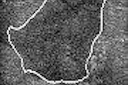
Ultrasound is moderately accurate in the diagnosis of substantial fatty atrophy of the supraspinatus or infraspinatus muscle, according to research published in Radiology.
"Although MR imaging remains the standard of reference for assessment of fatty atrophy of the rotator cuff muscles, the evaluation of supraspinatus and infraspinatus muscle belly should be included when shoulder US is performed in patients suspected of having rotator cuff tears," wrote a research team from the University Hospital Zurich in Zurich, Switzerland.
To prospectively evaluate the accuracy of ultrasound in depicting fatty atrophy of the supraspinatus (SSP) and infraspinatus (ISP) muscles, the team studied 65 consecutive patients with possible rotator cuff tears (Radiology, September 28, 2005). Ultrasound was performed immediately before and after MR arthrography by one of two musculoskeletal radiologists using a multifrequency linear probe (7-9 MHz) on an Elegra scanner (Siemens Medical Solutions, Malvern, PA).
Tissue harmonic imaging was used on all patients, and each muscle belly was evaluated in two planes. Images were then stored on a PACS network (Image Devices, Idstein, Germany) for later review.
At a later date, the two radiologists -- blinded to patient data and imaging parameters -- later evaluated the visibility of muscle contour, pennate pattern, central tendon, and muscle echogenicity. MR was used as the reference standard.
The accuracy, sensitivity, and specificity for ultrasound in diagnosing fatty atrophy in the supraspinatus and infraspinatus muscles for both readers are shown below, along with the readers' rates of overestimating and underestimating the grade of atrophy for the muscles.
|
|||||||||||||||||||||||||||||||||||
The study team found that the two readers more commonly overestimated the grade of supraspinatus muscle atrophy than they underestimated it.
The study team found that interobserver agreement for the supraspinatus muscle was moderate (k = 0.55), while agreement for the infraspinatus muscle was substantial (k = 0.71).
The authors acknowledged some study limitations, including the basing the accuracy of ultrasound evaluations on a selection of static images, which may be inferior compared with clinical routine when exams are performed dynamically. In addition, the relatively small proportion of patients with severe fatty atrophy may have reduced the diagnostic accuracy of ultrasound, the authors stated.
By Erik L. Ridley
AuntMinnie.com staff writer
October 19, 2005
Related Reading
No slam dunk: Partial cuff tears defy MRI, US diagnosis, June 17, 2005
Enthesopathy on shoulder MRI may tag severity of rotator cuff tears, February 24, 2005
Acromion evaluation proves tricky for x-rays and MRI, February 21, 2005
Standard and extremity MR bear brunt of shoulder imaging, November 17, 2004
Copyright © 2005 AuntMinnie.com


![Examples of ultrasound findings and techniques. (A) Images in a 39-year-old male patient with a mass in the left thigh. The mass is heterogeneous on the B-mode US image (compared with the patient in D) and showed increased microvascularity (superb microvascular imaging [SMI]) and shear-wave elastography (SWE) values. Undifferentiated pleomorphic sarcoma was diagnosed at biopsy (with pleomorphic rhabdomyosarcoma in surgical specimen). (B) Images in an 18-year-old male patient with a mass in the left leg. The mass is hypoechoic on the B-mode image, with no other findings suggestive of malignancy. The lesion is in contact with the cortex of the tibia, which is slightly irregular. CT revealed a doubtful anteromedial tibial erosion. The microvascular study demonstrated high vascularization, suggestive of malignancy. Periosteal Ewing sarcoma was diagnosed with both histologic and immunohistochemical confirmation. (C) Images in a 69-year-old female patient with a lump growing on the outside of the left leg. Multiple SWE examinations were performed (please note the high values obtained in the measurements, whereas the color map highlights the stiffness relative to adjacent tissues). SMI showed areas of increased vascularization to target for sampling. Undifferentiated spindle cell sarcoma was diagnosed at biopsy, with residual leiomyosarcoma in the surgical specimen after neoadjuvant therapy. (D) Images in a 56-year-old female patient with a mass in the right thigh. The mass is heterogeneous at both B-mode ultrasound (similar to patient A) and MRI (coronal T2-weighted spectral attenuated inversion recovery [SPAIR]; T1-weighted pre-contrast and postcontrast imaging), which even shows uptake after the administration of paramagnetic contrast material, which is traditionally suggestive of malignancy. Low values at SMI and elastography are suggestive of benignity. Spindle cell lipoma was diagnosed at biopsy, with atypical spindle cell lipomatous tumor in the surgical specimen.](https://img.auntminnie.com/mindful/smg/workspaces/default/uploads/2026/08/images-radiol250278fig2.APCFLSvX6p.jpg?auto=format%2Ccompress&fit=crop&h=100&q=70&w=100)


![Examples of ultrasound findings and techniques. (A) Images in a 39-year-old male patient with a mass in the left thigh. The mass is heterogeneous on the B-mode US image (compared with the patient in D) and showed increased microvascularity (superb microvascular imaging [SMI]) and shear-wave elastography (SWE) values. Undifferentiated pleomorphic sarcoma was diagnosed at biopsy (with pleomorphic rhabdomyosarcoma in surgical specimen). (B) Images in an 18-year-old male patient with a mass in the left leg. The mass is hypoechoic on the B-mode image, with no other findings suggestive of malignancy. The lesion is in contact with the cortex of the tibia, which is slightly irregular. CT revealed a doubtful anteromedial tibial erosion. The microvascular study demonstrated high vascularization, suggestive of malignancy. Periosteal Ewing sarcoma was diagnosed with both histologic and immunohistochemical confirmation. (C) Images in a 69-year-old female patient with a lump growing on the outside of the left leg. Multiple SWE examinations were performed (please note the high values obtained in the measurements, whereas the color map highlights the stiffness relative to adjacent tissues). SMI showed areas of increased vascularization to target for sampling. Undifferentiated spindle cell sarcoma was diagnosed at biopsy, with residual leiomyosarcoma in the surgical specimen after neoadjuvant therapy. (D) Images in a 56-year-old female patient with a mass in the right thigh. The mass is heterogeneous at both B-mode ultrasound (similar to patient A) and MRI (coronal T2-weighted spectral attenuated inversion recovery [SPAIR]; T1-weighted pre-contrast and postcontrast imaging), which even shows uptake after the administration of paramagnetic contrast material, which is traditionally suggestive of malignancy. Low values at SMI and elastography are suggestive of benignity. Spindle cell lipoma was diagnosed at biopsy, with atypical spindle cell lipomatous tumor in the surgical specimen.](https://img.auntminnie.com/mindful/smg/workspaces/default/uploads/2026/08/images-radiol250278fig2.APCFLSvX6p.jpg?auto=format%2Ccompress&fit=crop&h=112&q=70&w=112)