Korean and Taiwanese physicians have proposed imaging breast elasticity with ultrasound, in combination with computer-aided analysis. They found that continuous ultrasound images, obtained during compression, could accurately classify benign and malignant lesions.
"Elastography is an imaging technique that depicts tissue stiffness by means of imaging tissue strain from external compression.... A complex radiofrequency extraction subsystem, however, is required to apply this technique to current (ultrasound) scanners," wrote Dr. Woo Kyung Moon and colleagues from Seoul National University Hospital and the Institute of Radiation Medicine in Seoul; National Chung Cheng University in Chiaya, Taiwan; and Changhua Christian Hospital in Changhua, Taiwan (Radiology, August 2005, Vol. 236:2, pp. 458-464).
For this study, a database was mined to produce 100 consecutive pathologically proven solid breast tumors obtained with sonographic probe compression. There were 60 benign tumors and 40 invasive carcinomas. Images were obtained with a real-time B-mode scanner with a 5-10 MHz transducer (Voluson 530, Kretz Technik, Zipf, Austria).
The tumor contours were segmented from the continuous ultrasound images, then three values for features of strain were computed: contour difference, shift distance, and area difference. Solidity was used as the feature for shape feature. A support vector machine was used to classify solid breast tumors based on these values.
![]() |
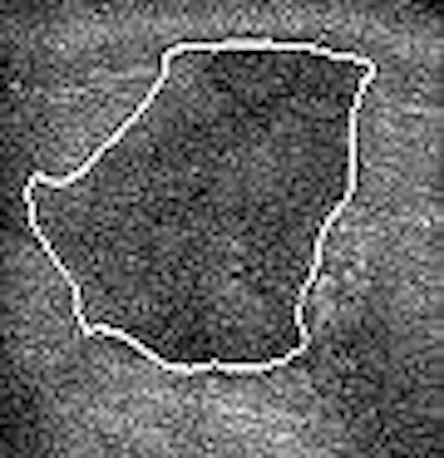
| Transverse continuous US images (a-h) of a malignant breast lesion. The white outline is the computer-delineated margin. In this case, the values for the three features of strain -- contour difference, shift difference, and area difference -- and the value for the one feature of shape (solidity) were 1.20%, 1.24%, 0.85%, and 3.69%, respectively. Figure 4, Moon WK, Chang R-F, Chen C-J, Chen D-R, Chen W-L. Solid Breast Masses: Classification with Computer-aided Analysis of Continuous US Images Obtained with Probe Compression. Radiology 2005;236:458-464. |
The results showed that the mean value of contour difference in malignant tumors was 3.52% on continuous ultrasound images. For shift distance the mean value was 2.62%, and for area difference it was 1.08%. For benign tumors the mean value of contour difference was 9.72%. The mean value for shift distance was 5.04%, and for area difference it was 3.17%. The accuracy for continuous ultrasound using the support vector machine was 87%, sensitivity was 85%, specificity was 88%, positive predictive value (PPV) was 83%, and negative predictive value (NPV) was 90%.
On noncontinuous ultrasound images, the mean value for contour difference in malignant tumors was 22.13%, for shift distance the mean value was 2.86%, and for area difference it was 6.18%. In benign tumors the mean value of contour difference was 56.23%. For shift difference the mean value was 5.4%, and for area difference it was 17.95%. Noncontinuous ultrasound with the support vector machine turned in an accuracy of 80%, sensitivity of 75%, specificity of 83%, PPV of 75%, and NPV of 83%.
The area under the receiver operating characteristic curve (Az) values for the three features of strain were significant higher than the Az value for the shape feature, the authors reported.
"The results of our study show the proposed method that is based on image segmentation and analysis of features of strain with continuous B-mode images can be used successfully to classify benign and malignant tumors," they stated.
The researchers cautioned that operating technique is important element in the success of this method. The ultrasonographer must use constant pressure on the scanning probe to avoid an inclined scanning probe and potentially having the tumor slip out of the scanning plane, they explained. Both practice and a compression plate can used to improve performance.
By Shalmali Pal
AuntMinnie.com staff writer
September 7, 2005
Related Reading
Ultrasound helps find occult breast cancer postsurgery, May 4, 2005
Hormonal variations can affect breast vascularity, US reveals, January 13, 2005
Tumor type, breast profile determine value of mammo, US, and MR, January 6, 2005
Copyright © 2005 AuntMinnie.com




![Examples of ultrasound findings and techniques. (A) Images in a 39-year-old male patient with a mass in the left thigh. The mass is heterogeneous on the B-mode US image (compared with the patient in D) and showed increased microvascularity (superb microvascular imaging [SMI]) and shear-wave elastography (SWE) values. Undifferentiated pleomorphic sarcoma was diagnosed at biopsy (with pleomorphic rhabdomyosarcoma in surgical specimen). (B) Images in an 18-year-old male patient with a mass in the left leg. The mass is hypoechoic on the B-mode image, with no other findings suggestive of malignancy. The lesion is in contact with the cortex of the tibia, which is slightly irregular. CT revealed a doubtful anteromedial tibial erosion. The microvascular study demonstrated high vascularization, suggestive of malignancy. Periosteal Ewing sarcoma was diagnosed with both histologic and immunohistochemical confirmation. (C) Images in a 69-year-old female patient with a lump growing on the outside of the left leg. Multiple SWE examinations were performed (please note the high values obtained in the measurements, whereas the color map highlights the stiffness relative to adjacent tissues). SMI showed areas of increased vascularization to target for sampling. Undifferentiated spindle cell sarcoma was diagnosed at biopsy, with residual leiomyosarcoma in the surgical specimen after neoadjuvant therapy. (D) Images in a 56-year-old female patient with a mass in the right thigh. The mass is heterogeneous at both B-mode ultrasound (similar to patient A) and MRI (coronal T2-weighted spectral attenuated inversion recovery [SPAIR]; T1-weighted pre-contrast and postcontrast imaging), which even shows uptake after the administration of paramagnetic contrast material, which is traditionally suggestive of malignancy. Low values at SMI and elastography are suggestive of benignity. Spindle cell lipoma was diagnosed at biopsy, with atypical spindle cell lipomatous tumor in the surgical specimen.](https://img.auntminnie.com/mindful/smg/workspaces/default/uploads/2026/08/images-radiol250278fig2.APCFLSvX6p.jpg?auto=format%2Ccompress&fit=crop&h=100&q=70&w=100)


![Examples of ultrasound findings and techniques. (A) Images in a 39-year-old male patient with a mass in the left thigh. The mass is heterogeneous on the B-mode US image (compared with the patient in D) and showed increased microvascularity (superb microvascular imaging [SMI]) and shear-wave elastography (SWE) values. Undifferentiated pleomorphic sarcoma was diagnosed at biopsy (with pleomorphic rhabdomyosarcoma in surgical specimen). (B) Images in an 18-year-old male patient with a mass in the left leg. The mass is hypoechoic on the B-mode image, with no other findings suggestive of malignancy. The lesion is in contact with the cortex of the tibia, which is slightly irregular. CT revealed a doubtful anteromedial tibial erosion. The microvascular study demonstrated high vascularization, suggestive of malignancy. Periosteal Ewing sarcoma was diagnosed with both histologic and immunohistochemical confirmation. (C) Images in a 69-year-old female patient with a lump growing on the outside of the left leg. Multiple SWE examinations were performed (please note the high values obtained in the measurements, whereas the color map highlights the stiffness relative to adjacent tissues). SMI showed areas of increased vascularization to target for sampling. Undifferentiated spindle cell sarcoma was diagnosed at biopsy, with residual leiomyosarcoma in the surgical specimen after neoadjuvant therapy. (D) Images in a 56-year-old female patient with a mass in the right thigh. The mass is heterogeneous at both B-mode ultrasound (similar to patient A) and MRI (coronal T2-weighted spectral attenuated inversion recovery [SPAIR]; T1-weighted pre-contrast and postcontrast imaging), which even shows uptake after the administration of paramagnetic contrast material, which is traditionally suggestive of malignancy. Low values at SMI and elastography are suggestive of benignity. Spindle cell lipoma was diagnosed at biopsy, with atypical spindle cell lipomatous tumor in the surgical specimen.](https://img.auntminnie.com/mindful/smg/workspaces/default/uploads/2026/08/images-radiol250278fig2.APCFLSvX6p.jpg?auto=format%2Ccompress&fit=crop&h=112&q=70&w=112)