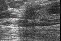
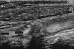
(Ultrasound Review) The sonographic findings in focal fibrocystic changes of the breast were described in a study recently published in Ultrasound Quarterly. Researchers at Baylor College of Medicine and Woman’s Hospital of Texas studied 58 patients that had 60 lesions with a pathologic diagnosis of fibrocystic changes (FC). The authors explained "an adequate understanding of the sonographic appearance of benign processes in the breast is essential to reduce the rate of repeat intervention resulting from discordance between imaging findings and histopathologic findings."
According to the authors, FC is defined as "a condition in which there are palpable lumps in the breast, usually associated with pain and tenderness, that fluctuate with the menstrual cycle and that become progressively worse until menopause. Histologically, FC includes macrocysts, microcysts, adenosis, apocrine change, fibrosis, or ductal hyperplasia."
Most patients underwent surgical biopsy after preoperative mammographic localization, three had core biopsy and one patient had fine-needle aspiration (FNA). On ultrasound, focal FC appeared as a solid mass in 47%, as heterogeneous echogenic tissue in 15%, as cysts in 13%, and in 25% of cases was not visible on ultrasound.
The criteria developed by Stavros were used to characterize benign, indeterminate or probably malignant. Of the 28 masses that were shown, 46% were classified indeterminate (neither malignant nor benign).
"In 23 patients, there was a localized palpable abnormality and fifteen of these had a sonographically visible mass or cyst," they reported. There were fifteen FC lesions that were not visible on ultrasound. Thirteen lesions were not shown mammographically, mostly due to dense breast tissue.
Only 38%of the lesions that were histologically proven to be focal FC were palpable. The authors reported that aside from fibroadenomas and gross cysts, FC fails to demonstrate a discreet mass. Also, they warned that a significant number of focal FC appears as solid masses.
"Sonographic findings in focal fibrocystic changes of the breast"M Shetty et al
Dept of radiology, Baylor College of Medicine and Woman’s Hospital of Texas, Houston,TX
Ultrasound Quarterly 2002 March; 18:35-40
By Ultrasound Review
May 29, 2002
Copyright © 2002 AuntMinnie.com

![Examples of ultrasound findings and techniques. (A) Images in a 39-year-old male patient with a mass in the left thigh. The mass is heterogeneous on the B-mode US image (compared with the patient in D) and showed increased microvascularity (superb microvascular imaging [SMI]) and shear-wave elastography (SWE) values. Undifferentiated pleomorphic sarcoma was diagnosed at biopsy (with pleomorphic rhabdomyosarcoma in surgical specimen). (B) Images in an 18-year-old male patient with a mass in the left leg. The mass is hypoechoic on the B-mode image, with no other findings suggestive of malignancy. The lesion is in contact with the cortex of the tibia, which is slightly irregular. CT revealed a doubtful anteromedial tibial erosion. The microvascular study demonstrated high vascularization, suggestive of malignancy. Periosteal Ewing sarcoma was diagnosed with both histologic and immunohistochemical confirmation. (C) Images in a 69-year-old female patient with a lump growing on the outside of the left leg. Multiple SWE examinations were performed (please note the high values obtained in the measurements, whereas the color map highlights the stiffness relative to adjacent tissues). SMI showed areas of increased vascularization to target for sampling. Undifferentiated spindle cell sarcoma was diagnosed at biopsy, with residual leiomyosarcoma in the surgical specimen after neoadjuvant therapy. (D) Images in a 56-year-old female patient with a mass in the right thigh. The mass is heterogeneous at both B-mode ultrasound (similar to patient A) and MRI (coronal T2-weighted spectral attenuated inversion recovery [SPAIR]; T1-weighted pre-contrast and postcontrast imaging), which even shows uptake after the administration of paramagnetic contrast material, which is traditionally suggestive of malignancy. Low values at SMI and elastography are suggestive of benignity. Spindle cell lipoma was diagnosed at biopsy, with atypical spindle cell lipomatous tumor in the surgical specimen.](https://img.auntminnie.com/mindful/smg/workspaces/default/uploads/2026/08/images-radiol250278fig2.APCFLSvX6p.jpg?auto=format%2Ccompress&fit=crop&h=100&q=70&w=100)


![Examples of ultrasound findings and techniques. (A) Images in a 39-year-old male patient with a mass in the left thigh. The mass is heterogeneous on the B-mode US image (compared with the patient in D) and showed increased microvascularity (superb microvascular imaging [SMI]) and shear-wave elastography (SWE) values. Undifferentiated pleomorphic sarcoma was diagnosed at biopsy (with pleomorphic rhabdomyosarcoma in surgical specimen). (B) Images in an 18-year-old male patient with a mass in the left leg. The mass is hypoechoic on the B-mode image, with no other findings suggestive of malignancy. The lesion is in contact with the cortex of the tibia, which is slightly irregular. CT revealed a doubtful anteromedial tibial erosion. The microvascular study demonstrated high vascularization, suggestive of malignancy. Periosteal Ewing sarcoma was diagnosed with both histologic and immunohistochemical confirmation. (C) Images in a 69-year-old female patient with a lump growing on the outside of the left leg. Multiple SWE examinations were performed (please note the high values obtained in the measurements, whereas the color map highlights the stiffness relative to adjacent tissues). SMI showed areas of increased vascularization to target for sampling. Undifferentiated spindle cell sarcoma was diagnosed at biopsy, with residual leiomyosarcoma in the surgical specimen after neoadjuvant therapy. (D) Images in a 56-year-old female patient with a mass in the right thigh. The mass is heterogeneous at both B-mode ultrasound (similar to patient A) and MRI (coronal T2-weighted spectral attenuated inversion recovery [SPAIR]; T1-weighted pre-contrast and postcontrast imaging), which even shows uptake after the administration of paramagnetic contrast material, which is traditionally suggestive of malignancy. Low values at SMI and elastography are suggestive of benignity. Spindle cell lipoma was diagnosed at biopsy, with atypical spindle cell lipomatous tumor in the surgical specimen.](https://img.auntminnie.com/mindful/smg/workspaces/default/uploads/2026/08/images-radiol250278fig2.APCFLSvX6p.jpg?auto=format%2Ccompress&fit=crop&h=112&q=70&w=112)