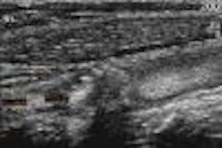
DENVER - Using a high-resolution, 10-minute ultrasound exam to study the anatomic structures of the wrist may help surgeons improve the outcome of carpal tunnel procedures and avoid complications. In a poster presentation at the American Academy of Neurology conference on Tuesday, investigators from Wake Forest University in Winston-Salem, NC, discussed the advantages of high-resolution ultrasound over current diagnostic techniques.
The gold standard for diagnosing carpal tunnel syndrome is a nerve conduction study, but "it is not perfect...it doesn't provide any anatomical information about the wrist, and it can be uncomfortable," explained presenter Michael Cartwright, a fourth-year medical student at Wake Forest. Dr. Ethan Wiesler, a hand-surgery specialist, was Cartwright’s co-author.
Carpal tunnel syndrome may also be diagnosed using MRI or CT, but those modalities can be expensive and time-consuming, Cartwright said.
Using an ATL HDI 5000 scanner (Philips Ultrasound, Bothell, WA) equipped with a 5.12 MHz transverse linear-array probe, the researchers imaged the wrists of 43 healthy volunteers, and 30 wrists of 16 patients with carpal tunnel syndrome.
The ultrasound sagittal cut -- placing the probe in the center of the palm -- displayed the median nerve, the flexor tendons, transverse carpal ligament, and bone boundaries of the tunnel, Cartwright said. In a cross-sectional view, the same structures could be visualized by placing the probe across the wrist at the base of the palm.
"We did the study to determine if we could see differences between normal and patient wrists, and one thing that we found is that the median nerve is greatly enlarged among patients, possibly due to inflammation," Cartwright said.
In the normal wrist, the nerve area measures about 8.8 square mm, he said, but in the subjects with carpal tunnel syndrome, the nerve area averaged 19.1 square mm. In some patients, ultrasound was able to visualize blood vessels, such as a small artery, close to the nerve and tendon.
"I think this diagnostic tool will be very helpful to the surgeons performing carpal tunnel release procedures," commented Dr. Todd Troost, chairman of the department of neurology at Wake Forest. "Surgeons can now have images to guide them in these operations. It may prevent complications of the surgery."
Dr. Ladislao Olivares, a neurologist at Hospital Medica Sur in Mexico City, Mexico, said that most patients with an early diagnosis of carpal tunnel syndrome could be treated with physical rehabilitation. But "once I decide to send them to surgery, this type of imaging will help because it provides a great deal of information we can't get now with standard studies of the wrist," he said.
By Edward SusmanAuntMinnie.com contributing writer
April 17, 2002
Copyright © 2002 AuntMinnie.com
![Examples of ultrasound findings and techniques. (A) Images in a 39-year-old male patient with a mass in the left thigh. The mass is heterogeneous on the B-mode US image (compared with the patient in D) and showed increased microvascularity (superb microvascular imaging [SMI]) and shear-wave elastography (SWE) values. Undifferentiated pleomorphic sarcoma was diagnosed at biopsy (with pleomorphic rhabdomyosarcoma in surgical specimen). (B) Images in an 18-year-old male patient with a mass in the left leg. The mass is hypoechoic on the B-mode image, with no other findings suggestive of malignancy. The lesion is in contact with the cortex of the tibia, which is slightly irregular. CT revealed a doubtful anteromedial tibial erosion. The microvascular study demonstrated high vascularization, suggestive of malignancy. Periosteal Ewing sarcoma was diagnosed with both histologic and immunohistochemical confirmation. (C) Images in a 69-year-old female patient with a lump growing on the outside of the left leg. Multiple SWE examinations were performed (please note the high values obtained in the measurements, whereas the color map highlights the stiffness relative to adjacent tissues). SMI showed areas of increased vascularization to target for sampling. Undifferentiated spindle cell sarcoma was diagnosed at biopsy, with residual leiomyosarcoma in the surgical specimen after neoadjuvant therapy. (D) Images in a 56-year-old female patient with a mass in the right thigh. The mass is heterogeneous at both B-mode ultrasound (similar to patient A) and MRI (coronal T2-weighted spectral attenuated inversion recovery [SPAIR]; T1-weighted pre-contrast and postcontrast imaging), which even shows uptake after the administration of paramagnetic contrast material, which is traditionally suggestive of malignancy. Low values at SMI and elastography are suggestive of benignity. Spindle cell lipoma was diagnosed at biopsy, with atypical spindle cell lipomatous tumor in the surgical specimen.](https://img.auntminnie.com/mindful/smg/workspaces/default/uploads/2026/08/images-radiol250278fig2.APCFLSvX6p.jpg?auto=format%2Ccompress&fit=crop&h=100&q=70&w=100)


![Examples of ultrasound findings and techniques. (A) Images in a 39-year-old male patient with a mass in the left thigh. The mass is heterogeneous on the B-mode US image (compared with the patient in D) and showed increased microvascularity (superb microvascular imaging [SMI]) and shear-wave elastography (SWE) values. Undifferentiated pleomorphic sarcoma was diagnosed at biopsy (with pleomorphic rhabdomyosarcoma in surgical specimen). (B) Images in an 18-year-old male patient with a mass in the left leg. The mass is hypoechoic on the B-mode image, with no other findings suggestive of malignancy. The lesion is in contact with the cortex of the tibia, which is slightly irregular. CT revealed a doubtful anteromedial tibial erosion. The microvascular study demonstrated high vascularization, suggestive of malignancy. Periosteal Ewing sarcoma was diagnosed with both histologic and immunohistochemical confirmation. (C) Images in a 69-year-old female patient with a lump growing on the outside of the left leg. Multiple SWE examinations were performed (please note the high values obtained in the measurements, whereas the color map highlights the stiffness relative to adjacent tissues). SMI showed areas of increased vascularization to target for sampling. Undifferentiated spindle cell sarcoma was diagnosed at biopsy, with residual leiomyosarcoma in the surgical specimen after neoadjuvant therapy. (D) Images in a 56-year-old female patient with a mass in the right thigh. The mass is heterogeneous at both B-mode ultrasound (similar to patient A) and MRI (coronal T2-weighted spectral attenuated inversion recovery [SPAIR]; T1-weighted pre-contrast and postcontrast imaging), which even shows uptake after the administration of paramagnetic contrast material, which is traditionally suggestive of malignancy. Low values at SMI and elastography are suggestive of benignity. Spindle cell lipoma was diagnosed at biopsy, with atypical spindle cell lipomatous tumor in the surgical specimen.](https://img.auntminnie.com/mindful/smg/workspaces/default/uploads/2026/08/images-radiol250278fig2.APCFLSvX6p.jpg?auto=format%2Ccompress&fit=crop&h=112&q=70&w=112)