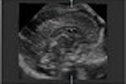
(Ultrasound Review) Prenatal researchers from St. Joannsspital in Salzburg, Austria, have compared nuchal translucency (NT) measurement using 2-D and 3-D ultrasound.
"The risk for trisomies increases with increasing maternal age and NT," they wrote. Improving measurement accuracy should increase screening sensitivity. The study had three objectives. The first was to assess the feasibility of 3-D NT measurement, secondly to determine whether 3-D was accurate and thirdly to compare 2-D and 3-D results. Their findings were published in the December issue of Ultrasound in Obstetrics and Gynecology.
Comparing 2-D and 3-D ultrasound measurements of nuchal translucency, the researchers found that 3-D ultrasound enabled a correct measurement more often. The difference between the two modes however was not statistically significant.
Two hundred and twenty-nine women with a fetus of crown rump length 45–84 mm were included for study. The mean of three nuchal translucency measurements was calculated using both 2-D and 3-D in the transverse and midsagittal planes. Examination duration was limited to twenty minutes and the mean examination time for 2-D and 3-D was comparable. There was a high correlation between 2-D and 3-D measurements and between midsagittal and transverse measurements. Abnormal cases also underwent 3-D surface reconstruction but this failed to add further diagnostic information.
In 60% of cases, three measurements were possible using 3-D, compared with 51.7% of 2-D cases. A successful measurement was obtained using 3-D in 98.6% of cases compared with 96.8% of cases using 2-D. All transvaginal examinations enabled successful measurement, but the majority (95%) of examinations were performed transabdominally.
Differentiation between the skin line and the amnion proved to be the biggest challenge for transabdominal 2-D examinations. Fetal was the main pitfall in obtaining a measurement using 3-D ultrasound.
"The possibility of rotating a stored volume and inspecting it in three orthogonal planes makes 3-D ultrasound a useful tool for nuchal translucency measurements, especially in doubtful cases," they wrote.
"Comparison between 2-D and 3-D ultrasound measurements of nuchal translucency"G Clementschitsch et al
Dept of Obstetrics and Gynecology, Prenatal Diagnosis and Therapy Unit, St Johannsspital Salzburg, Austria
Ultrasound Obstet Gynecol 2001(December); 18:475-480
By Ultrasound Review
January 4, 2002
Copyright © 2002 AuntMinnie.com
![Examples of ultrasound findings and techniques. (A) Images in a 39-year-old male patient with a mass in the left thigh. The mass is heterogeneous on the B-mode US image (compared with the patient in D) and showed increased microvascularity (superb microvascular imaging [SMI]) and shear-wave elastography (SWE) values. Undifferentiated pleomorphic sarcoma was diagnosed at biopsy (with pleomorphic rhabdomyosarcoma in surgical specimen). (B) Images in an 18-year-old male patient with a mass in the left leg. The mass is hypoechoic on the B-mode image, with no other findings suggestive of malignancy. The lesion is in contact with the cortex of the tibia, which is slightly irregular. CT revealed a doubtful anteromedial tibial erosion. The microvascular study demonstrated high vascularization, suggestive of malignancy. Periosteal Ewing sarcoma was diagnosed with both histologic and immunohistochemical confirmation. (C) Images in a 69-year-old female patient with a lump growing on the outside of the left leg. Multiple SWE examinations were performed (please note the high values obtained in the measurements, whereas the color map highlights the stiffness relative to adjacent tissues). SMI showed areas of increased vascularization to target for sampling. Undifferentiated spindle cell sarcoma was diagnosed at biopsy, with residual leiomyosarcoma in the surgical specimen after neoadjuvant therapy. (D) Images in a 56-year-old female patient with a mass in the right thigh. The mass is heterogeneous at both B-mode ultrasound (similar to patient A) and MRI (coronal T2-weighted spectral attenuated inversion recovery [SPAIR]; T1-weighted pre-contrast and postcontrast imaging), which even shows uptake after the administration of paramagnetic contrast material, which is traditionally suggestive of malignancy. Low values at SMI and elastography are suggestive of benignity. Spindle cell lipoma was diagnosed at biopsy, with atypical spindle cell lipomatous tumor in the surgical specimen.](https://img.auntminnie.com/mindful/smg/workspaces/default/uploads/2026/08/images-radiol250278fig2.APCFLSvX6p.jpg?auto=format%2Ccompress&fit=crop&h=100&q=70&w=100)


![Examples of ultrasound findings and techniques. (A) Images in a 39-year-old male patient with a mass in the left thigh. The mass is heterogeneous on the B-mode US image (compared with the patient in D) and showed increased microvascularity (superb microvascular imaging [SMI]) and shear-wave elastography (SWE) values. Undifferentiated pleomorphic sarcoma was diagnosed at biopsy (with pleomorphic rhabdomyosarcoma in surgical specimen). (B) Images in an 18-year-old male patient with a mass in the left leg. The mass is hypoechoic on the B-mode image, with no other findings suggestive of malignancy. The lesion is in contact with the cortex of the tibia, which is slightly irregular. CT revealed a doubtful anteromedial tibial erosion. The microvascular study demonstrated high vascularization, suggestive of malignancy. Periosteal Ewing sarcoma was diagnosed with both histologic and immunohistochemical confirmation. (C) Images in a 69-year-old female patient with a lump growing on the outside of the left leg. Multiple SWE examinations were performed (please note the high values obtained in the measurements, whereas the color map highlights the stiffness relative to adjacent tissues). SMI showed areas of increased vascularization to target for sampling. Undifferentiated spindle cell sarcoma was diagnosed at biopsy, with residual leiomyosarcoma in the surgical specimen after neoadjuvant therapy. (D) Images in a 56-year-old female patient with a mass in the right thigh. The mass is heterogeneous at both B-mode ultrasound (similar to patient A) and MRI (coronal T2-weighted spectral attenuated inversion recovery [SPAIR]; T1-weighted pre-contrast and postcontrast imaging), which even shows uptake after the administration of paramagnetic contrast material, which is traditionally suggestive of malignancy. Low values at SMI and elastography are suggestive of benignity. Spindle cell lipoma was diagnosed at biopsy, with atypical spindle cell lipomatous tumor in the surgical specimen.](https://img.auntminnie.com/mindful/smg/workspaces/default/uploads/2026/08/images-radiol250278fig2.APCFLSvX6p.jpg?auto=format%2Ccompress&fit=crop&h=112&q=70&w=112)