(Ultrasound Review) - This research, published in the August issue of the Journal of Ultrasound in Medicine, is presented by radiologists from Rome and relates to the high-resolution ultrasound examination of the lacrimal glands in women with Sjögren syndrome.
This chronic autoimmune disease affects the lacrimal and salivary glands, causing swelling in the early stages, as well as fibrosis and fat deposition. In its final stages, the glands shrink.
This study examined 15 women with the disease and 15 volunteers, employing high-resolution ultrasound to visualize the lacrimal glands. The glands were visible bilaterally on ultrasound in six patients, and all of these had an abnormal Schirmer test and xerophthalmia.
Of the nine patients whose glands were not visible, only two had an abnormal Schirmer test, and four had xerophthalmia.
"The lacrimal gland lies in the superior, anterior, and lateral part of the orbit and is composed of two subglands, called orbital and palpebral parts, divided by the musculus levator palpebrae superioris," the researchers wrote.
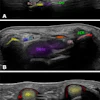
In the normal subjects, ultrasound showed the lacrimal gland, as a small hypoechoic area in front of the distal part of the lacrimal artery, and the lacrimal artery was located along the posterior border of the orbit. On ultrasound, the lacrimal gland was barely distinguishable from the surrounding orbital fat.
Ultrasound was performed using either a 7.5–10 MHz or 10–13 MHz linear-array transducer. The lacrimal artery was visualized with power Doppler in all cases, in both patients and volunteers, and the waveform reflected a low impedance flow pattern. For patients with Sjögren syndrome, the mean resistive index of the lacrimal artery was significantly higher (0.72) than in the normal subjects (0.61).
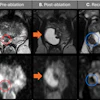
"In conclusion, sonography is certainly able to confirm the clinical suggestion of lacrimal involvement in Sjögren syndrome, to quantify the degree of activity and therefore to predict the possibility for an anti-inflammatory therapy (visibility of the glands means presence of vital glandular parenchyma that can be saved by therapy), and to detect abnormal swelling of the glands that might express lymphoma," The researchers wrote. "The diagnostic role of MRI may be limited to the differentiation of normal-sized glands from atrophic glands when glands are invisible with ultrasonography."
J Ultrasound Med 2000; 19:505–509
Francesco Giovagnorio et al
Via del Pettinari 40, 00186 Rome, Italy
By Ultrasound Review
October 12, 2000