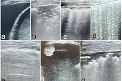
High-resolution ultrasound may be used for quickly assessing forearm lacerations, according to research published June 8 in Injury.
A team led by Umesh Kumar, PhD, from Banaras Hindu University in Varanasi, Uttar Pradesh, India, found that the modality achieved near-perfect performance for evaluating tendon injuries. It also achieved high marks for finding nerve and arterial injuries while keeping diagnosis times under 10 minutes on average.
“This study supports its [high-resolution ultrasound] integration into routine trauma care to improve diagnostic accuracy and better outcomes,” the Kumar team wrote.
Forearm lacerations are often tied to tendon, nerve, and vessel injuries. While clinical exams are frequently performed for these injuries, they may not be enough, the researchers noted. Accurate and quick diagnosis is needed to avoid any potential functional impairment.
Previous studies suggest that high-resolution ultrasound can address this issue. Kumar and colleagues investigated just how effective the modality is in diagnosing tendon, nerve, and vascular injuries in acute trauma cases.
For the prospective observational study, the researchers included 38 patients with forearm lacerations between 2022 and 2024. All patients underwent a clinical exam followed by high-resolution ultrasound ranging from 3 MHz to 15 MHz. The researchers used surgical exploration as the gold standard for their study.
The Kumar team reported the following findings:
Ultrasound achieved a diagnostic accuracy of 99.6% in tendon injuries, including a sensitivity of 98.6% and a specificity of 100%.
Ultrasound achieved an accuracy of 96.5% for finding nerve injuries.
Ultrasound achieved an accuracy of 90.7% for identifying arterial injuries.
Ultrasound-based diagnosis in forearm lacerations led to an average time of 9.53 minutes.
The researchers also highlighted that the outcome of evaluation with high-resolution ultrasound with clinical examination was better than that of isolated clinical examination for tendon and neurovascular injuries.
The results support high-resolution ultrasound’s integration into standard protocols, the study authors wrote. This includes serving as an adjunct diagnostic tool to evaluate lacerations in emergency settings.
“It provides rapid, accurate, and comprehensive diagnosis, and helps to streamline the surgical planning and execution,” the authors added.
The team called for future studies to have larger sample sizes and to take place at different centers for further validation.
The full study can be accessed here.