MRI helps clinicians assess the neural involvement in endometriosis and could help them prevent irreversible nerve damage and chronic sensorimotor neuropathy in women suffering from the condition, Cleveland Clinic researchers have reported.
MRI findings regarding the state of pelvic nerves involved in endometriosis are key to planning treatment, wrote a group led by Ceylan Colak, MD. The team's review of MRI's role for this indication was published January 3 in RadioGraphics.
"Evidence of irreversible damage can … be seen at MRI, and radiologists should evaluate pelvic nerves that are commonly involved in endometriosis in their search pattern and report template to ensure that this information is incorporated into treatment planning," Colak and colleagues noted.
Endometriosis most commonly manifests in the pelvis, and its most common symptom is pain, they explained. Nerves in the pelvis can become entrapped in endometriosis, including the sciatic, obturator, femoral, pudendal, and inferior hypogastric nerves and the inferior hypogastric and lumbosacral plexuses, and this neural involvement can present not only as pain but also as weakness, numbness, incontinence, and even paraplegia.
The condition is often treated with laparoscopic excision and identifying any neural structures involved can help with surgical planning, the team explained. Endovaginal ultrasound is also used to "map" endometriosis lesions, but MRI offers the field of view and spatial resolution that produce reliable images of the pelvic nerves, particularly with techniques such as high-spatial-resolution anatomic imaging -- including three-dimensional isotropic imaging -- and contrast-enhanced 3D short inversion time inversion-recovery (STIR) fast spin-echo sequences.
"MRI has excellent soft-tissue resolution and can demonstrate the fascicular pattern, signal intensity, and thickness of nerves," the group wrote.
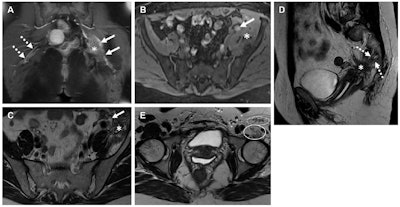
 Spectrum of imaging features of nerves involved in endometriosis in three women. (A) Coronal T1W contrast-enhanced MR image in a 44-year-old woman shows a thickened left sciatic nerve with increased enhancement (solid arrows) and a fibrotic mass (*). The right-side sciatic nerve is normal in appearance (dashed arrows). (B-D) Axial T1W image (without contrast enhancement) with fat saturation (B) in a 43-year-old woman shows a cystic endometriotic lesion with signal hyperintensity (arrow in B). The adjacent left iliacus muscle is asymmetrically thickened due to involvement by endometriosis (* in B). Axial T2-weighted MR image (C) at the same level as B shows T2 shading in the cystic lesion (arrow in C) and a fibrotic mass replacing the iliacus muscle (* in C). Sagittal T2-weighted MR image (D) shows the sacral nerve roots (dashed arrows in D) encased by a fibrotic endometriotic mass (* in D). (E) Axial T2-weighted MR image in a 37-year-old woman shows atrophy of the left iliopsoas (solid oval) and sartorius muscles (dashed oval), both innervated by the femoral nerve. Images and caption courtesy of the RSNA.
The bottom line? MRI may improve patient care when it comes to assessing endometriosis, especially since the involvement of pelvic nerves in the condition may be "underrecognized and underreported," according to Colak and colleagues.
"Early identification of nerve involvement in endometriosis is important for patient counseling, treatment planning, and determining the surgical approach," they concluded. "Radiologists who interpret pelvic endometriosis studies should identify the pelvic nerves involved in endometriosis and report this information to the clinician for treatment planning and improved patient outcomes."
The complete study can be found here.


