Can synthetic MRI replace conventional exams for evaluating brain health? It appears so, according to research published February 15 in Acta Radiologica.
Synthetic MRI's benefits could translate into better patient care, wrote a team led by Mengsha Zou, PhD, of Sun Yat-sen University in Gangzhou, China.
"Synthetic magnetic resonance imaging might replace ... conventional MR sequences in brain evaluation to shorten scan time and obtain multiple quantitative parameters," the group noted.
MRI is a go-to modality for many indications, producing high-resolution images of soft tissue, the investigators explained. But scan times can be long -- which can complicate care for children or patients who are claustrophobic.
Synthetic MRI images are generated by multiple-delay-multiple-echo (MDME) sequences that create a range of up to 10 different contrast images and three quantitative maps (T1, T2, and proton density); the technique shortens scanning time and offers more cost-effective access to MRI data, as conventional MRI requires obtaining multiple contrast-weighted images separately, the team explained.
"Considering these advantages, the possibility of replacing conventional MRI sequences with MDME sequence-driving synthetic MRI in brain evaluation to shorten scan times and obtain multiple quantitative parameters is worth considering," the authors noted.
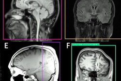
Zou and colleagues sought to compare synthetic and conventional MR images in a study that included 200 patients ranging in age from newborns to seniors; all study participants underwent both conventional and synthetic MRI imaging between January and December 2020. Synthetic images were generated from MDME data using a software package called SyMRI (SyntheticMR, Linköping, Sweden).
The investigators categorized patients into two groups: a healthy control cohort (118) and one with abnormal MRI findings (82). Two radiologists compared the images, and the researchers analyzed image quality, artifacts, and diagnostic performance of each set.
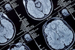
They found that synthetic MRI images showed equal to or greater than signal-to-noise and contrast-to-noise ratios in all study participants; in patients 2 years old and younger, synthetic T2-fluid-attenuated inversion recovery imaging showed significantly higher cerebellum gray/white matter contrast-to-noise ratio. The group also noted that on synthetic T1-weighted imaging, most study participants (90.7%) showed high signal intensity within the superior sagittal sinus. The investigators did not find any differences between synthetic and conventional MRI for lesion diagnosis.
"[Compared] with conventional MRI sequences, multiple-delay-multiple-echo sequence-derived synthetic MRI showed a shorter scan time, significantly higher or comparable signal-to-noise ratio and contrast-to-noise ratio, and good imaging quality in a comprehensive analysis of brain structures in a large and multi-age sample," they concluded.
The study did have some limitations, however, one of which being a lack of reader experience in interpreting synthetic MR images and another the presence of artifacts such as hyperintensity in the superior sagittal sinus on T1-weighted images, according to the authors.
"[The] possibility of high signal intensity within the superior sagittal sinus on synthetic T1-weighted images should be considered to avoid misdiagnosing thrombus," they noted.
In any case, synthetic MRI shows promise for improving patient care, according to the researchers.
"These results pave the way for the clinical utility of synthetic MRI in brain evaluation," they wrote.