Functional MRI (fMRI) can help detect which young adults may be at increased risk for depression or anxiety after stressful events, according to a study from Duke University in the February 4 issue of Neuron.
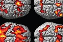
The researchers found that increased activity in the amygdala region of the brain predicted greater symptoms of depression and anxiety in response to future stressful events as much as one to four years in the future. The findings could lead to early interventions for those who need help, according to lead author Dr. Johnna Swartz, a psychology and neuroscience postdoctoral associate, and colleagues.
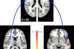
The researchers measured activity in the amygdala in 750 college students between the ages of 18 and 22 years. All subjects said they had no previous history of depression or anxiety disorders at the start of the study (Neuron, February 4, 2015, Vol. 85:3, pp. 505-511).
The participants were shown photos of angry or fearful faces while they underwent fMRI. After the scans, the students were contacted by email every three months and asked to complete an online survey regarding their current mood and any stressful life events. Approximately 350 students responded, and more than half of them completed an assessment at least one year after scanning.
Participants whose scans recorded higher activity in the amygdala in response to the photos went on to assess themselves as more prone to depression or anxiety after stressful events during the follow-up surveys, Swartz and colleagues found.
Along with identifying a risk marker for developing future mental health symptoms, the results suggest that finding therapies or drugs that decrease the activity of the amygdala may be most effective for preventing or alleviating stress-related depression and anxiety.